Etiology
Acquired torticollis is usually idiopathic. While most patients with acquired torticollis do not present with an overt pattern of heritability, one comparative study found that 8.7% of patients have a family history of dystonia (a figure much higher than the background prevalence).[5]
This phenomenon suggests that there is a genetic predisposition in some cases.[6][7]
Genetic defects on chromosome 8 (dystonia 6, or DYT-6) and chromosome 18 (dystonia 7, DYT-7) have been associated with adult-onset disease, but do not account for most cases.[8]
A genome-wide association study did not find a common prevalent genetic cause.[9]
In some patients there is a clear temporal link to trauma involving the neck, but the role of trauma as an etiologic or triggering factor remains unclear.[10] Acquired torticollis can occur as an acute or delayed (tardive) complication of therapy with dopamine receptor-blocking drugs, such as antipsychotics or other dopamine antagonists (e.g., metoclopramide, prochlorperazine). Tardive dystonia is estimated to occur in 3% of patients treated with long-term antipsychotics.[11] Cervical dystonia can occur as a symptomatic manifestation of multiple disease processes that cause dysfunction of the basal ganglia. These include perinatal asphyxia/cerebral palsy, Huntington disease, and Wilson disease.
Pathophysiology
While it is conventional that dystonia is a disorder of the brain, the pathophysiology of acquired torticollis remains poorly understood. The observable end result is a centrally-activated (but involuntary) contraction of skeletal muscle, resulting in abnormal posture.
Neurophysiologic and functional imaging studies have implicated abnormalities in the basal ganglia, as well as sensory and motor cortices. An imaging study demonstrated differences in a functional network between basal ganglia and sensorimotor cortex areas that could be modulated by botulinum toxin therapy.[12] There is also evidence of abnormalities in dopamine signaling, sensory processing, cortical motor inhibition, and sensorimotor integration.[8][13] Quantitative voxel-based morphometry studies have shown decreased volumes of caudate, putamen, and sensorimotor cortex compared with control subjects.[14]
Classification
Clinical classification by type
Clinically, acquired torticollis is classified by three criteria:
Primary head position, which is defined specifically as torticollis (neck rotation), anterocollis (neck flexion), retrocollis (neck extension), laterocollis (lateral neck flexion), sagittal shift, or lateral shift. Combinations of these types frequently occur.
Presence or absence of more widespread dystonia, such as focal (restricted to muscles of neck and shoulders), segmental (involving >1 contiguous body area), multifocal (involving noncontiguous body areas), or generalized.
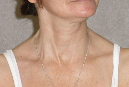
Primary (idiopathic) or secondary (e.g., due to antipsychotic drugs, other dopamine antagonists, cerebral palsy, Wilson disease, Huntington disease).[Figure caption and citation for the preceding image starts]: Patient with moderate left torticollis or neck rotation, mild right laterocollis or lateral neck flexion, and mild retrocollis or neck extensionFrom the collection of Dr David Sommer [Citation ends].
 [Figure caption and citation for the preceding image starts]: Patient with severe left torticollis (note hypertrophy of right sternocleidomastoid muscle)From the collection of Dr David Sommer [Citation ends].
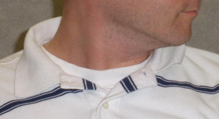
Use of this content is subject to our disclaimer