Case history
Case history #1
A 29-year-old woman presents with a 3-month history of worsening headaches and increasing visual loss. She describes occasional episodes of bilateral visual grayouts lasting 20 seconds that may be precipitated by bending forward or standing. Over the last 2 weeks she has often heard a "whooshing" sound in both ears, synchronous with her pulse, that is more noticeable when she is about to go to sleep. Her visual acuity is 20/30 (6/9 meters) in each eye. Fundus examination shows bilateral optic disk swelling, and Humphrey automated perimetry shows enlargement of the blind spot and scattered abnormal test locations. Magnetic resonance imaging shows a partially empty sella, and a magnetic resonance venogram shows no evidence of a thrombosis but does demonstrate bilateral transverse sinus venous stenoses. Lumbar puncture opening pressure is 280 mm H2O. [Figure caption and citation for the preceding image starts]: Bilateral disk edemaFrom the personal collection of Dr M. Wall; used with permission [Citation ends].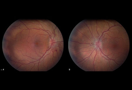 [Figure caption and citation for the preceding image starts]: Magnetic resonance image (MRI) of empty sella on sagittal viewFrom the personal collection of Dr M. Wall; used with permission [Citation ends].
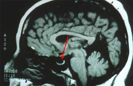
Case history #2
A 42-year-old man presents with a 2-month history of increasing migraine headaches that are worse in the morning and associated with nausea. He has occasional episodes of transient visual loss in both eyes that last 30 seconds before recovering. He has been overweight for many years and admits to gaining weight over the last couple of years. His body mass index at the time of presentation is 34.
Other presentations
Other common presentations include severe headaches and visual loss in young women. Usually the course is chronic.
Use of this content is subject to our disclaimer