Images and videos
Images


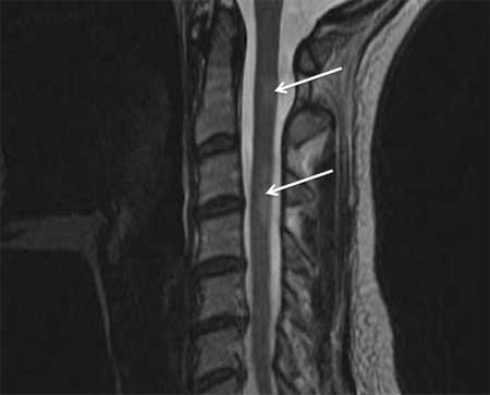
Transverse myelitis
Sagittal T2-weighted MRI showing longitudinally extensive TM lesion
From the personal collection of Dean M. Wingerchuk, MD, MSc, FRCP(C); used with permission
See this image in context in the following section/s:

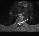
Transverse myelitis
Axial T2-weighted MRI of cervical spinal cord showing myelitis lesion
From the personal collection of Dean M. Wingerchuk, MD, MSc, FRCP(C); used with permission
See this image in context in the following section/s:
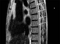
Transverse myelitis
Sagittal T2-weighted MRI showing multiple sclerosis-related myelitis lesion
From the personal collection of Dean M. Wingerchuk, MD, MSc, FRCP(C); used with permission
See this image in context in the following section/s:

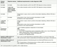
Transverse myelitis
The McDonald criteria for the diagnosis of multiple sclerosis
Created using data from Thompson AJ, et al. Lancet Neurol 2018;17:162–73
See this image in context in the following section/s:

Transverse myelitis
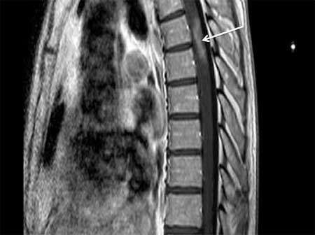
Sagittal T2-weighted MRI of cervical spinal cord showing myelitis lesion
From the personal collection of Dean M. Wingerchuk, MD, MSc, FRCP(C); used with permission
See this image in context in the following section/s:
Transverse myelitis
Sagittal T1-weighted MRI showing enhancement of neuromyelitis optica-related lesion
From the personal collection of Dean M. Wingerchuk, MD, MSc, FRCP(C); used with permission
See this image in context in the following section/s:
Use of this content is subject to our disclaimer