Criteria
Transverse Myelitis Consortium Working Group[1][23]
Inclusion criteria:
Development of sensory, motor, or autonomic dysfunction attributable to the spinal cord
Bilateral signs and/or symptoms (though not necessarily symmetric)
Clearly defined sensory level
Exclusion of extra-axial compressive etiology by neuroimaging (magnetic resonance imaging [MRI] or computed tomography [CT] myelography)
Inflammation of the spinal cord demonstrated by cerebrospinal pleocytosis, elevated IgG index, or MRI gadolinium enhancement
Progression to nadir between 4 hours and 21 days following the onset of symptoms.
Exclusion criteria:
History of previous radiation to the spine within the last 10 years
Clear arterial distribution consistent with thrombosis of the anterior spinal artery
Abnormal flow voids on the spinal cord surface consistent with dural-based arteriovenous fistula
Serologic or clinical evidence of connective tissue disease
Central nervous system manifestations of syphilis, Lyme disease, HIV, human T-lymphotropic virus-1, Mycoplasma, other viral infection
Brain MRI abnormalities suggestive of multiple sclerosis (MS)
History of clinically apparent optic neuritis.
Criteria for diagnosis of neuromyelitis optica spectrum disorder (NMOSD)[74]
Core clinical characteristics
Optic neuritis
Acute myelitis
Area postrema syndrome: episode of otherwise unexplained hiccups or nausea and vomiting
Acute brain stem syndrome
Symptomatic narcolepsy or acute diencephalic clinical syndrome with NMOSD-typical diencephalic MRI lesions
Symptomatic cerebral syndrome with NMOSD-typical brain lesions
Diagnostic criteria for NMOSD with anti-aquaporin-4 IgG (AQP4-IgG)
At least 1 core clinical characteristic
Positive test for AQP4-IgG using best available detection method (cell-based assay strongly recommended)
Exclusion of alternative diagnoses
Diagnostic criteria for NMOSD without AQP4-IgG or NMOSD with unknown AQP4-IgG status
At least 2 core clinical characteristics occurring as a result of one or more clinical attacks and meeting all of the following requirements:
At least 1 core clinical characteristic must be optic neuritis, acute myelitis with longitudinally extensive TM (LETM), or area postrema syndrome
Dissemination in space (2 or more different core clinical characteristics)
Fulfillment of additional MRI requirements, as applicable
Negative test(s) for AQP4-IgG using best available detection method, or testing unavailable
Exclusion of alternative diagnoses
Additional MRI requirements for NMOSD without AQP4-IgG and NMOSD with unknown AQP4-IgG status
Acute optic neuritis: requires brain MRI showing a) normal findings or only nonspecific white matter lesions; or b) optic nerve MRI with T2-hyperintense lesion or T1-weighted gadolinium-enhancing lesion extending over >1/2 optic nerve length or involving optic chiasm
Acute myelitis: requires associated intramedullary MRI lesion extending over >3 contiguous segments (LETM) or >3 contiguous segments of focal spinal cord atrophy in patients with prior history compatible with acute myelitis
Area postrema syndrome: requires associated dorsal medulla/area postrema lesions
Acute brain stem syndrome: requires associated peri-ependymal brain stem lesions
The 2017 revised McDonald diagnostic criteria for MS[75]
[Figure caption and citation for the preceding image starts]: The McDonald criteria for the diagnosis of multiple sclerosisCreated using data from Thompson AJ, et al. Lancet Neurol 2018;17:162–73 [Citation ends].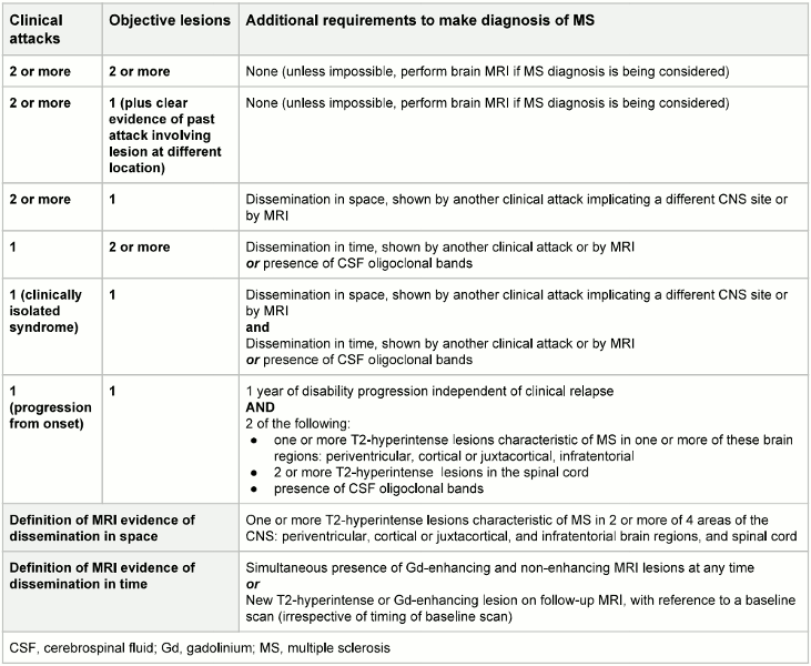
Use of this content is subject to our disclaimer