Differentials
Mucous cyst of the vestibule
SIGNS / SYMPTOMS
Tends to be soft, <2 cm in diameter, superficial, and smooth.[18]
INVESTIGATIONS
Diagnosis is clinical.
Vulval hematoma
Vulval fibroma
Vulval lipoma
SIGNS / SYMPTOMS
Skin-colored, soft, fatty tumor of the subcutaneous tissue; typically asymptomatic and slow growing.[20][Figure caption and citation for the preceding image starts]: Vulval lipomaFrom the personal collection of Colleen Kennedy Stockdale [Citation ends].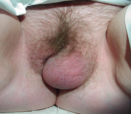
Usually in the labia majora, and further lipomas may be found on lower abdomen or thighs.[18]
INVESTIGATIONS
Diagnosis is clinical.
Cyst of the canal of Nuck
SIGNS / SYMPTOMS
Cystic swelling in the inguinal crease or anterior labia majora. Not crossed by the labium minus.
Arises from remnants of peritoneum as it passes through the inguinal canal, so cysts may occur anywhere along the path of the inguinal canal or within the labia majora.
INVESTIGATIONS
Diagnosis is clinical.
Epidermal inclusion cyst (sebaceous, keratinous, or epidermoid cyst)
Malignant lesion of Bartholin gland
SIGNS / SYMPTOMS
Tends to present in older women (>50 years) as an irregular nodular vulval mass, with or without ulcerations.[3][12][Figure caption and citation for the preceding image starts]: Squamous cell cancerFrom the personal collection of Colleen Kennedy Stockdale [Citation ends].
INVESTIGATIONS
Biopsy of the lesion confirms or excludes malignancy.
Use of this content is subject to our disclaimer