History and exam
Key diagnostic factors
common
malar (butterfly) rash
The characteristic malar or butterfly rash occurs in 30% to 40% of patients, and may be more common in female patients.[40][75]
Most commonly erythema over the cheeks and bridge of nose, sparing the nasolabial folds.
Malar rash often recurs after sun exposure. Recent onset of photosensitivity is supportive of the diagnosis.
[Figure caption and citation for the preceding image starts]: a) Photograph of a face with skin rashes sparing the bridge of the nose and malar area. b) Photograph of a face showing asymmetric hyperpigmented, polycylic, and annular scaly plaques with scaling involving pre-auricular area and cheekRajasekharan C et al. BMJ Case Reports. 2013;2013:bcr-2012-007886 [Citation ends].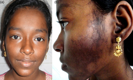 [Figure caption and citation for the preceding image starts]: Malar rash: butterfly shape, flat, non-tender erythematosus rash over the cheek and noseKumar N et al. BMJ Case Reports. 2013;2013:bcr-2012-008101 [Citation ends].
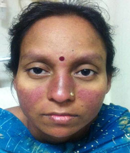
photosensitive rash
Rash occurs after sun exposure. It can be painful and pruritic and usually lasts a few days, healing without scarring.
discoid rash
Erythematous raised patches with adherent keratotic scaling and follicular plugging.
Atrophic scarring may occur in older lesions.
Other diagnostic factors
common
fatigue
weight loss
Often parallels the course of the illness.
Vomiting and diarrhea may contribute to weight loss. SLE is associated with an increased risk for cancer, and this should be considered as a potential cause of weight loss.
fever
Unexplained fever is common and characteristic for SLE.[1] It is thought to represent active disease.
Elevated C-reactive protein is suggestive of infection rather than underlying disease.
Exclusion of infection is important before initiating immunosuppressive therapy in a patient with SLE to prevent reactivation or exacerbation of chronic infection.[50]
oral ulcers
Occur in up to 45% of patients.[52]
Typically painless but prolonged and recurrent.
alopecia
Hair thinning and patchy alopecia are an understandable concern in young women with SLE. Parallels the systemic disease course.
Usually nonscarring.
Areas of scarring alopecia are more characteristic of chronic discoid lupus.
arthralgia/arthritis
Arthralgia is common in SLE. Inflammatory joint symptoms occur in >50% of patients.[50][53][76]
The arthritis can be similar to rheumatoid arthritis, although classically nonerosive.
Monoarthritis of a large joint is unusual in a patient with SLE and should initially prompt the search for another cause such as infection or avascular necrosis.
fibromyalgia
Poorly localized symmetrical musculoskeletal pain with no diurnal variation.
Poorly responsive to analgesics/nonsteroidal anti-inflammatory drugs suggests coexisting fibromyalgia; typical tender points should be checked for.
Raynaud phenomenon
Color changes of the digits induced by cold or emotion. Typical triphasic color change from white to blue to red in fingers and/or toes. Invariably bilateral and occurs in as many as 50% of patients at disease onset, though often predating other features of SLE.[50][53] It is often less severe than that seen in systemic sclerosis.
Raynaud phenomenon leading to ulceration is unusual and should prompt consideration of other causes.
chest pain and shortness of breath
Pleuritis is more common than pericarditis. In a survey of 1000 European patients with SLE, pleuritis and/or pericarditis was noted in 16%.[77] Pleuritis can be either unilateral or bilateral. In a minority, pleural effusions can coexist.
Other cardiovascular manifestations include myocarditis, endocarditis, venous or arterial thrombosis, and premature atheromatous coronary artery disease.
Shrinking lung syndrome is a rare manifestation of SLE characterized by shortness of breath, chest pain, a raised hemidiaphragm, and a restrictive pattern on pulmonary function tests.[62]
venous or arterial thrombosis
The presence of antiphospholipid antibodies increases the risk of venous or arterial thrombosis.[78]
hypertension
May occur as part of cardiopulmonary manifestations.
Renal involvement is usually subclinical and usually develops in the first few years of illness. Hypertension may be one of the first signs of lupus nephritis. Blood pressure and urinalysis looking for proteinuria and hematuria should be routinely performed.
signs of nephrosis (e.g., edema)
Renal involvement is present in approximately 50% to 70% of patients, and may be more common in male patients.[53][40] Renal involvement is usually subclinical and usually develops in the first few years of illness.
Blood pressure and urinalysis looking for proteinuria and hematuria should be routinely performed.
lymphadenopathy
Peripheral lymphadenopathy is more often regional than generalized. The nodes are usually nontender, vary in size from shotty (clusters of small lymph nodes, each a few mm) to 3 to 4 cm, and often are in the cervical and axillary regions.
Hilar lymphadenopathy is uncommon.
Patients with lymphadenopathy are more likely to have constitutional manifestations.
Lymphoma and infectious mononucleosis should be excluded.
Histology of lymph node biopsies in SLE frequently shows reactive hyperplasia.
abdominal pain, vomiting, or diarrhea
Occurs as part of gastrointestinal manifestation of SLE. Caused by lupus peritonitis or mesenteric artery occlusion. Peritonitis is rare.
uncommon
nose ulcers
Typically painless but prolonged and recurrent.
poorly localized proximal limb inflammatory pain with weakness
Suggestive of an associated myositis; if present, creatinine phosphokinase will be elevated.
dysrhythmias (e.g., tachycardia), conduction defects, or unexplained cardiomegaly
Myocarditis should be suspected in these patients.
CNS signs: seizures, cranial nerve abnormalities, cognitive defects, psychosis
Major central nervous system (CNS) involvement in SLE is uncommon.[54] Other possible causes should be excluded.
Antiribosomal P is significantly associated with CNS involvement and psychosis.[71][72] Standardization of antiribosomal P assays is required.
Investigations in patients with neuropsychiatric symptoms should be similar to that of the general population presenting with the same symptoms.[47]
dysphagia
Occurs as part of gastrointestinal manifestation of SLE.
Due to esophageal hypomotility.
Risk factors
strong
female sex
The incidence of SLE is higher in women than in men; reported gender ratios range from 2:1 to 15:1.[8][9][10]
The increased frequency of SLE among women has been associated with the effects of estrogen.[6][13]
Alopecia, photosensitivity, oral ulcers, arthritis, and malar rash may be more common among female patients.[40]
age >30 years
African descent in Europe and US
The US Centres for Disease Control and Prevention National Lupus Registries found that prevalence was higher in African Americans (230.9 per 100,000 and 26.7 per 100,000 for women and for men, respectively), than in Hispanics (120.7 per 100,000 and 18.0 per 100,000, respectively) or white populations (84.7 per 100,000 and 8.9 per 100,000, respectively).[11]
Reported incidences in Africa are low (0.3 per 100,000 person-years).[6] This may reflect under-diagnosis due to resource shortage.
drugs
Clinical and serologic manifestations can occur in patients taking some medications.[25][41]
The first reported association was with procainamide, but other commonly implicated drugs include minocycline, terbinafine, sulfasalazine, isoniazid, phenytoin, and carbamazepine.[26][27][28][29][30][31][32][33]
Symptoms of drug-induced lupus erythematosus resolve when the offending drug is discontinued.
Some of the reported associations between drug use and a subsequent diagnosis of SLE may be due to protopathic bias (when a treatment is inadvertently prescribed for an early manifestation of a disease that has not yet been diagnosed).[41]
weak
sun exposure
Exposure to ultraviolet (UV) radiation can exacerbate skin lesions in lupus erythematosus patients (photosensitivity). Sun exposure is the most obvious environmental factor that exacerbates SLE.[42]
Prospective, methodologically robust studies are required to evaluate the relationship between UV-B and incident SLE.
family history of SLE
Use of this content is subject to our disclaimer