Approach
There are no internationally validated diagnostic criteria for SLE.
In a validation cohort, the 2019 European League Against Rheumatism/American College of Rheumatology (ACR) classification criteria had a sensitivity of 96.1% and specificity of 93.4%.[1] Comparable figures for the ACR 1997 criteria were 82.8% sensitivity and 93.4% specificity, and for the Systemic Lupus International Collaborating Clinics 2012 criteria were 96.7% sensitivity and 83.7% specificity.[1]
Constitutional symptoms and signs
Fatigue, fever, and weight loss are common symptoms that occur at some time during the course of the disease.
Fatigue is common, occurring in 80% to 100% of patients, but it does not correlate with disease activity.[48][49] Fatigue from other causes, such as anemia, hypothyroidism, medications (e.g., beta-blockers), depression, fibromyalgia, and social stresses, should be considered.
Unexplained fever is common and characteristic for SLE.[1] It is thought to represent active disease. Exclusion of infection is important before initiating immunosuppressive therapy in a patient with SLE to prevent reactivation or exacerbation of chronic infection.[50] Fever persisting despite treatment with a nonsteroidal anti-inflammatory drug or acetaminophen should raise suspicion for an infectious or drug-related etiology. All patients presenting with persistent fever should have an appropriate symptom-targeted infection screen.
Weight loss in SLE may be related to disease activity or its treatment. Patients with SLE may have esophageal hypomotility leading to dysphagia. Vomiting and diarrhea may contribute to weight loss. SLE is associated with an increased risk for cancer, and this should be considered as a potential cause of weight loss.
Lymphadenopathy
Peripheral lymphadenopathy is more often regional than generalized. The nodes are usually nontender, vary in size from shotty (clusters of small lymph nodes, each a few mm) to 3 to 4 cm, and are often in the cervical and axillary regions. Hilar lymphadenopathy is uncommon. Patients with lymphadenopathy are more likely to have constitutional manifestations. Lymphoma and infectious mononucleosis should be excluded. Histology of lymph node biopsies in SLE frequently shows reactive hyperplasia.
Mucocutaneous symptoms and signs
Skin manifestations are a common presentation of SLE.
The characteristic malar or butterfly rash occurs in 30% to 40% of patients, and may be more common in female patients.[40][51] This erythematous rash extends from the cheeks over the bridge of the nose, sparing the nasolabial folds. It can be painful and pruritic, usually lasts for a few days, and heals without scarring. Malar rash often recurs after sun exposure. When rash occurs both above and below the neck it is referred to as acute generalized cutaneous lupus rash. Recent onset of photosensitivity is supportive of the diagnosis.
[Figure caption and citation for the preceding image starts]: Malar rash: butterfly shape, flat, non-tender erythematosus rash over the cheek and noseKumar N et al. BMJ Case Reports. 2013;2013:bcr-2012-008101 [Citation ends].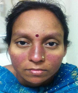
[Figure caption and citation for the preceding image starts]: a) Photograph of a face with skin rashes sparing the bridge of the nose and malar area. b) Photograph of a face showing asymmetric hyperpigmented, polycylic, and annular scaly plaques with scaling involving pre-auricular area and cheekRajasekharan C et al. BMJ Case Reports. 2013;2013:bcr-2012-007886 [Citation ends].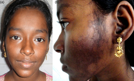
Other distinct categories of rash include discoid lupus, which presents as erythematous raised patches with adherent keratotic scaling and follicular plugging. Atrophic scarring may occur in older lesions. The latter patterns are less likely to be associated with systemic disease, but many patients are antinuclear antibody (ANA)-positive.
Mouth ulcers, and less frequently nose ulcers, occur in up to 45% of patients. They are usually large and painless, in contrast to herpetic lesions.[52] These ulcers often improve with simple local measures and their course parallels the disease course.
Active systemic disease can lead to diffuse patchy alopecia, which is reversible once disease is controlled. Discoid lesions leave permanent scarring alopecia.
Musculoskeletal symptoms and signs
Musculoskeletal symptoms are common, occurring in the majority of patients at some time during the course of the illness. Determining the distribution and the nature of the symptoms is helpful. A diurnal variation in pain, worse in the mornings with associated stiffness, suggests an underlying inflammatory component. Arthritis tends to be symmetrical and is typically nonerosive. Although uncommon, joint deformity may occur; joint deformity in the absence of erosive disease is referred to as Jaccoud arthritis. Correctable ulnar deviation and joint subluxations in the hands in the absence of radiologic damage are characteristic. Patients with SLE may also develop myositis leading to muscle weakness and pain.
A full history and musculoskeletal examination, looking for tenosynovitis and peripheral joint synovitis, particularly in the hands, should be performed. X-rays of affected joints should be requested. Septic arthritis should always be excluded in a patient presenting with a monoarthritis. The affected joint should be aspirated and fluid sent for microscopy and culture. Poorly localized proximal limb inflammatory pain with weakness may suggest an associated myositis and, if present, creatinine phosphokinase will be elevated. Diffuse musculoskeletal pain without a distinct diurnal variation may suggest coexisting fibromyalgia; typical tender points should be examined.
Raynaud phenomenon
Recent onset of triphasic color change in both hands and feet in a young woman due to exposure to cold or emotional stress should prompt a search for other features of SLE. The nail folds (for capillary nail fold changes) and peripheral pulses should be examined. The skin over the dorsum of the hands should be checked for sclerodactyly and features of systemic sclerosis or mixed connective tissue disease considered.
Renal symptoms and signs
Renal involvement is present in approximately 50% to 70% of patients, and may be more common in male patients.[40][53] Lupus nephritis is more common in Hispanic and black patients, and those with more severe disease in other organ systems. Those with antibodies to double-stranded (ds)DNA are more likely to develop glomerulonephritis. Most patients are asymptomatic. Other presentations include hypertension, nephrotic syndrome, or renal failure. Urinalysis may demonstrate the presence of hematuria, casts (red cell, granular, tubular, or mixed), or proteinuria.
Central nervous system symptoms and signs
Major central nervous system involvement in SLE is uncommon.[54] Seizures, cranial nerve abnormalities, and psychiatric illnesses are the most common. Cranial nerve abnormalities may manifest as visual field defects, blindness, papilledema, nystagmus, ptosis, or facial palsy. Myasthenia gravis and multiple sclerosis should be excluded. Psychiatric illnesses include psychosis, depressive disorders, and organic brain syndromes. Cerebral infarcts may occur and are usually related to coexisting positive antiphospholipid antibodies.
Case reports suggest that catatonia may be a manifestation of neuropsychiatric SLE, particularly in the presence of serologies and symptoms indicative of an active lupus flare.[55]
The diagnosis of cerebral involvement in SLE is clinical. Other causes such as sepsis, uremia, malignant hypertension, epilepsy, myasthenia gravis, and multiple sclerosis should be excluded.
Cardiopulmonary symptoms and signs
Cardiovascular manifestations of SLE include pericarditis, myocarditis, endocarditis, arterial and venous thrombosis, and premature atherosclerotic coronary artery disease. The risk of cardiovascular events (myocardial infarction and stroke) is two- to threefold higher in patients with SLE compared with the general population.[56][57][58][59][60]
Myocarditis should be suspected in patients with tachycardia, arrhythmias, conduction defects, or unexplained cardiomegaly. Nonbacterial Libman-Sacks endocarditis is uncommon.
Pulmonary manifestations of SLE include pleuritis, pleural effusions, diffuse interstitial lung disease, pulmonary hypertension and, rarely, pulmonary hemorrhage.[61] Pulmonary embolism should be excluded in patients with SLE presenting with pleuritic chest pain, dyspnea, and hemoptysis, particularly if antiphospholipid antibodies are positive. Pleural effusions in SLE are usually unilateral and generally exudative. Other causes of a pleural effusion should be excluded.
Shrinking lung syndrome is a rare respiratory manifestation of SLE characterized by dyspnea, chest pain, a raised hemidiaphragm, and a restrictive pattern on pulmonary function tests.[62]
Hematologic symptoms and signs
Anemia, leukopenia, and thrombocytopenia are common hematologic manifestations of SLE.[63] Anemia is usually secondary to chronic disease and improves with control of disease activity. Hemolytic anemia is not common, but can be very severe. Leukopenia is usually due to lymphopenia and to a lesser extent neutropenia. Thrombocytopenia is also frequently seen and other causes should be excluded. The presence of antiphospholipid antibodies increases the risk of venous and arterial thromboses.
Gastrointestinal symptoms and signs
SLE can affect any part of the gastrointestinal tract.[64] Oral ulcers are common.[65] Dysphagia is less common and is due to esophageal hypomotility. Abdominal pain, vomiting, and diarrhea may be caused by lupus peritonitis or mesenteric artery occlusion, but other causes of an acute abdomen should be excluded. Although rare, lupus peritonitis may mimic appendicitis. Pancreatitis may be due to SLE, but it is important to exclude treatment such as azathioprine as the underlying cause. Chronic active hepatitis may occur in SLE.
Serositis
Pleuritis and pericarditis is much more common than peritonitis. In the absence of any other explanation, the diagnosis of SLE should be considered in a patient with anterior chest pain suggestive of pleuritis and pericarditis (especially in at-risk patients such as women of reproductive age). The patient should be asked about mucocutaneous and musculoskeletal features, which, if present, may suggest the diagnosis.
Initial tests
Positive ANA is diagnostic of SLE when it occurs together with the ACR revised criteria for the classification of SLE.[1]
The following tests should be performed in anyone with suspected SLE:
Complete blood count and clotting screen: a prolongation of the partial thromboplastin time would suggest the presence of lupus anticoagulant and should prompt checking of antiphospholipid antibodies. Antiphospholipid antibodies should also be checked in patients with a history of recurrent spontaneous abortions and thromboses
Infection screen to include blood and urine cultures should be obtained in febrile patients. Septic arthritis should always be excluded in a patient presenting with a monoarthritis as it needs to be treated expeditiously
BUN and electrolytes to exclude or confirm possible renal involvement
Elevated erythrocyte sedimentation rate and CRP are suggestive of active disease, but infection must be excluded
Urinalysis should be done in all patients suspected of having SLE and regularly in patients with SLE, even in the absence of symptoms. All patients with lupus nephritis have proteinuria
Autoantibodies for antinuclear factor, dsDNA, and Smith antigen. A positive ANA in itself is not diagnostic as it may be positive in other connective tissue diseases such as rheumatoid arthritis, systemic sclerosis, Sjogren syndrome, thyroid disease, chronic infectious diseases, and inflammatory bowel disease, and in patients treated with certain drugs such as procainamide, hydralazine, isoniazid, and chlorpromazine. ANAs in a low titer also occur in healthy people. One in 3 will have a positive ANA at the screening dilution of 1:40 and 1 in 20 will have an ANA titer of 1:160.[66] As ANA can be positive in so many conditions, the result of a positive ANA has to be interpreted in the light of the clinical history and symptoms. Rarely, the ANA can be negative in SLE, especially in anti-Ro-antibody-positive lupus (Ro is also known as Sjogren syndrome A or Sjogren antibody). The ACR recommends the immunofluorescence ANA test using human epithelial type 2 (HEp-2) substrate as the gold standard for ANA testing.[67][68] Anti-dsDNA and anti-Smith antibodies are highly specific for SLE and often are confirmatory of the diagnosis, if present.[69][70] High titers of anti-dsDNA antibodies are markers of disease activity and high levels predict worse outcome in lupus nephritis.
Subsequent tests
Hematologic
Coombs test should be ordered if initial blood count shows anemia and features of hemolysis, such as elevated MCV and reticulocyte count.
Complement levels should be considered but are not necessary to diagnose SLE. They can be used in the setting of significant organ manifestations such as cerebritis or nephritis. Sequential rather than single measurements are necessary to be of value, in order to follow response to treatment or confirm worsening disease.
Immunologic
Antiphospholipid antibodies should be ordered in patients with a history of venous or arterial thromboses, miscarriages, or in patients with a prolonged activated PTT.
Skin biopsy is often not necessary to confirm the diagnosis of mucocutaneous manifestations as these are typically diagnosed clinically, but would be performed if diagnosis is in doubt. Skin biopsy of affected areas may show classic immune deposits at the dermal-epidermal junction on immunofluorescence or nonspecific inflammation.
Musculoskeletal
If there are symptoms and signs of musculoskeletal involvement, x-rays of affected joints should be requested.
If there is evidence of poorly localized proximal limb inflammatory pain with weakness, creatinine phosphokinase may be done to exclude myositis.
Renal
24-hour urine collection for protein or spot urine for protein/creatinine ratio should be performed if the urinalysis is abnormal.
A renal ultrasound should be performed in patients with abnormal urinary sediment.
A renal biopsy is the most sensitive and specific test for confirming the diagnosis of lupus nephritis and grading the extent of involvement by the International Society of Nephrology (ISN) and Renal Pathology Society (RPS) classification of lupus nephritis.[5] As renal involvement usually develops in the first few years of illness, blood pressure, urinalysis, and estimated glomerular filtration rate should be monitored. If any are abnormal, specialist opinion with a view to biopsy should be considered. If glomerulonephritis is present, it is classified on the basis of the ISN/RPS system.
Cerebral
Cerebral manifestations are typically diagnosed clinically. Central nervous system (CNS) lupus can be a diagnostic challenge. Brain MRI may be necessary if the diagnosis is in doubt and will show small focal areas of increased signal, which could be areas of inflammation. These lesions may resolve with treatment.
In patients with SLE with progressive cognitive loss, clinical evidence of SLE activity should be sought, and other causes (such as infection, electrolyte disturbance, vitamin or thyroid deficiency, or medication side-effects) need to be excluded.
Antiribosomal P is significantly associated with CNS involvement and psychosis.[71][72] Standardization of antiribosomal P assays is required.
Cardiopulmonary
Patients presenting with cardiopulmonary symptoms should have a chest x-ray and ECG done routinely.[73] Depending on the presenting complaint, echocardiogram, pulmonary function tests, or chest CT may be required. Patients presenting with pleural effusions need pleural aspiration to confirm the cause.

How to take a venous blood sample from the antecubital fossa using a vacuum needle.

How to record an ECG. Demonstrates placement of chest and limb electrodes.
Use of this content is subject to our disclaimer