Images and videos
Images


Frostbite
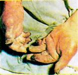
Grade IV frostbite injury inappropriately thawed using boiling water
Lloyd EL, BMJ 1994; 309:531-534
See this image in context in the following section/s:

Frostbite
Immersion foot (trench foot)
Lloyd EL, BMJ 1994; 309:531-534
See this image in context in the following section/s:
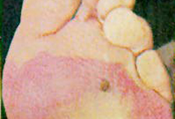
Frostbite
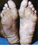
Grade II frostbite injury of the foot
Lloyd EL, BMJ 1994; 309:531-534
See this image in context in the following section/s:

Frostbite
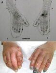
(A) Technetium-99m scans of the hands of a patient with frostbite. The terminal digits have reduced signal (especially in the left hand), suggesting that substantial tissue necrosis has occurred. (B) Clinical picture after a 5-day iloprost infusion showing the close correlation between the initial technetium-99m scans and the subsequent clinical appearance
Hallam M-J, BMJ 2010;341:c5864
See this image in context in the following section/s:

Frostbite
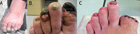
A typical frostbite affecting the hallux and third left toes showing the initial injury at presentation at base camp on Everest (A), at 6 weeks (B), and at 10 weeks (C). Note the delayed surgical amputation of the hallux after definitive demarcation and the recovery of the third digit after appropriate management
Hallam M-J, BMJ 2010;341:c5864
See this image in context in the following section/s:
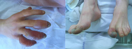
Frostbite
Typical frostbite injuries in the hands and feet of a climber with mildly haemorrhagic bullae presenting 3 days after exposure. The bullae were aseptically aspirated and a 5-day iloporost infusion resulted in a complete recovery
Hallam M-J, BMJ 2010;341:c5864
See this image in context in the following section/s:
Use of this content is subject to our disclaimer