Approach
The diagnosis is clinical, and additional investigations are only used to help determine the prognosis. Serial photographs can be helpful to document evolution of the injury and healing.[1]
History and examination
All patients have a history of exposure to freezing weather. Patients usually describe a sensation of cold in affected extremities, followed by loss of sensation or numbness. If rewarming has begun, patients may report extreme pain during or after the rewarming phase.[1][9][24][25][26][27]
Skin changes vary according to the severity of the injury. Erythema can occur but is more commonly seen with chilblains, a milder condition. True frostbite injuries appear similar at initial presentation, so severity classification should be applied after rewarming has occurred.[16]
The degree of severity is determined by the depth of the freezing and subsequent injury:[4]
First degree: purplish discoloration with a white or yellowish waxy discolouration in the area of injury, associated with numbness and erythema. Mild oedema is common, but there is no tissue loss.
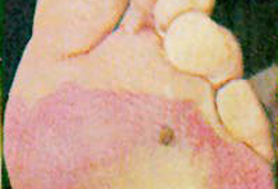
Second degree: superficial skin vesiculation with clear or milky fluid in blisters, surrounded by erythema and oedema. [Figure caption and citation for the preceding image starts]: Grade II frostbite injury of the footLloyd EL, BMJ 1994; 309:531-534 [Citation ends].
Third degree: may initially present as 2nd degree, but deep blisters characterised by purple, blood-containing fluid appear within 24 hours, indicating injury into the reticular dermis and beyond the dermal vascular plexus.
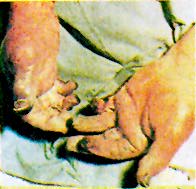
Fourth degree: injury is through the dermis, involving subcutaneous tissue, muscle, nerves, and bone. The tissue appears black and necrotic. Less severe bone injury in children may cause developmental digital deformities.[Figure caption and citation for the preceding image starts]: Grade IV frostbite injury inappropriately thawed using boiling waterLloyd EL, BMJ 1994; 309:531-534 [Citation ends].
The neurovascular status of the affected limb should be assessed (sensation, perfusion) and documented.
The pattern of bleb (blister) formation is a useful indicator of the severity of the injury. If blebs form distally all the way along the extremity, the injury is predominantly second degree. If blebs form proximally but not distally, injury to the distal tissue is third or fourth degree and there is a danger of tissue loss.[1][9][24][25][26][27] Patients may have fractures or joint dislocation due to weight-bearing on frostbitten feet, or concurrent trauma in the field. Signs of obvious dislocation or fracture (joint deformity, point tenderness) should be identified.
Imaging
Imaging is not required for the diagnosis of frostbite but can be used to assess the severity of the injury and determine the prognosis.
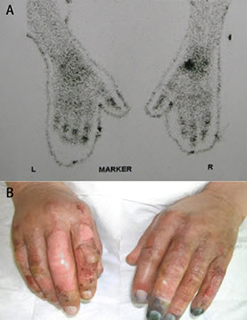
Technetium-99m pertechnetate scintigraphy provides an index of the perfusion of the affected extremity. It provides a sensitive and specific assessment of the extent of deep tissue and any bony injury. Some studies have found good correlation between scintigraphy findings at 48 hours and both intra-operative findings and ultimate prognosis.[2][3] Scintigraphy can also be used to assess response to therapy.[Figure caption and citation for the preceding image starts]: (A) Technetium-99m scans of the hands of a patient with frostbite. The terminal digits have reduced signal (especially in the left hand), suggesting that substantial tissue necrosis has occurred. (B) Clinical picture after a 5-day iloprost infusion showing the close correlation between the initial technetium-99m scans and the subsequent clinical appearanceHallam M-J, BMJ 2010;341:c5864 [Citation ends].
Early magnetic resonance angiography is an emerging modality and has the advantage of being more readily available. It may be useful to predict tissue viability and guide earlier surgical amputation.[4][28] It may also have a role in guiding therapies such as treatment with iloprost or recombinant tissue plasminogen activator.[4]
Thermography and duplex imagery are emerging modalities whose findings correlate with clinical outcome.[29] Their utility is yet to be determined.
Plain radiography will not determine frostbite severity but is required to assess for associated fractures and dislocations when suspected.
Use of this content is subject to our disclaimer