Aetiology
There is no specific identifiable causative factor; however, there is much speculation regarding aetiology.
Traumatic events may be recalled and linked to the ganglion in anywhere from 10% to 40% of patients.[7]
Scapholunate injury has also been hypothesised to lead to dorsal ganglia.[8]
Arthrographic studies have demonstrated radiopaque dye passing from the wrist joint into the ganglion cyst uni-directionally, leading to the theory that a small hole in the wrist capsule may cause a one-way valve allowing growth of the cyst.[9]
Ganglions may also represent benign tumours of synovial origin; however, there is no specific synovial lining noted on histological examination.[10]
Synovial fluid leaking into the surrounding tissue may lead to formation of a cyst wall enclosing cystic fluid.[11]
Mucoid degeneration of collagen in the surrounding tissue may lead to cyst formation.
A definitive link between ganglion cyst development and traumatic injury has not been established.
Pathophysiology
Ganglion cysts are usually small structures measuring 1 to 3 cm in diameter and located on the radial aspect. Impingement or surrounding of the radial artery can occur during cyst development. Occasionally, occult ganglions (<1 cm) or larger ganglions (up to 8 cm) have been reported. Cysts can be singular or multi-loculated and are usually located next to a joint or the surrounding tendons. Macroscopically, they tend to be smooth, white, and firm with an underlying stalk connection to the joint surface. They are mobile, non-adherent to underlying tissue, compressible, and usually not directly painful on palpation. The outer wall is comprised of randomly oriented collagen fibres with no definitive endothelial lining.[10] The cyst itself is filled with a thick, gelatinous, clear mucin comprised of glucosamine, globulin, hyaluronic acid, and albumin.[12]
Cysts may compress surrounding neurovascular structures and patients may experience wrist pain, paraesthesia, intrinsic muscle paralysis, and/or coolness of the hand or fingers as a result.
Dorsally located cysts are usually connected to the scapholunate interosseous ligament in the area of the dorsal capsular attachments; however, the stalk may be long enough to have the palpable mass located a distance away from the dorsoradial wrist. Two thirds of volar cysts are connected to the radioscaphoid joint and one third to the scaphotrapezial joint.[13]
There is no documented history of malignant transformation of a ganglion cyst.
Classification
Anatomic
Volar: between the flexor carpi radialis tendon and the radial artery.
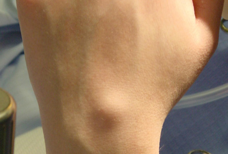
Dorsal: usually connected to the scapholunate ligament or scaphotrapeziotrapezoidal joint. [Figure caption and citation for the preceding image starts]: Typical dorsal wrist ganglion cystFrom the collection of Marco Rizzo, MD, Mayo Clinic; used with permission [Citation ends].
Occult: vague dorsal wrist pain with wrist flexion.
Use of this content is subject to our disclaimer