Clinical findings do not always correlate with symptoms: some patients are troubled by symptoms but have minimal defects on examination, whereas others are relatively free of symptoms but have significant defects. No single sign, symptom, or test result can establish the diagnosis of dry eye disease (DED).[2]Amescua G, Ahmad S, Cheung AY, et al. Dry eye syndrome preferred practice pattern®. Ophthalmology. 2024 Apr;131(4):P1-49.
https://www.doi.org/10.1016/j.ophtha.2023.12.041
http://www.ncbi.nlm.nih.gov/pubmed/38349301?tool=bestpractice.com
Diagnosis relies on a combination of detailed history taking, clear documentation, ophthalmologist-led examination, and specific diagnostic tests.[1]Hakim FE, Farooq AV. Dry eye disease: an update in 2022. JAMA. 2022 Feb 1;327(5):478-9.
http://www.ncbi.nlm.nih.gov/pubmed/35103781?tool=bestpractice.com
[3]Craig JP, Nichols KK, Akpek EK, et al. TFOS DEWS II definition and classification report. Ocul Surf. 2017 Jul;15(3):276-83.
https://www.sciencedirect.com/science/article/pii/S1542012417301192?via%3Dihub
http://www.ncbi.nlm.nih.gov/pubmed/28736335?tool=bestpractice.com
This should include evidence of systemic signs with referral to appropriate specialties if necessary.
History
The basic history should establish and describe:[1]Hakim FE, Farooq AV. Dry eye disease: an update in 2022. JAMA. 2022 Feb 1;327(5):478-9.
http://www.ncbi.nlm.nih.gov/pubmed/35103781?tool=bestpractice.com
[2]Amescua G, Ahmad S, Cheung AY, et al. Dry eye syndrome preferred practice pattern®. Ophthalmology. 2024 Apr;131(4):P1-49.
https://www.doi.org/10.1016/j.ophtha.2023.12.041
http://www.ncbi.nlm.nih.gov/pubmed/38349301?tool=bestpractice.com
[3]Craig JP, Nichols KK, Akpek EK, et al. TFOS DEWS II definition and classification report. Ocul Surf. 2017 Jul;15(3):276-83.
https://www.sciencedirect.com/science/article/pii/S1542012417301192?via%3Dihub
http://www.ncbi.nlm.nih.gov/pubmed/28736335?tool=bestpractice.com
Symptoms and signs (e.g., irritation, excessive tearing; burning, stinging, mild itching, dry, foreign body sensations; photophobia, blurry vision; contact lens intolerance, redness, mucous discharge, increased blinking, eye fatigue; symptoms that worsen throughout the day)
Exacerbating conditions (e.g., decreased humidity, fans, reading, digital device use)
Symptom duration.
A more detailed history should establish risk factors that can aid with diagnosis and management:[2]Amescua G, Ahmad S, Cheung AY, et al. Dry eye syndrome preferred practice pattern®. Ophthalmology. 2024 Apr;131(4):P1-49.
https://www.doi.org/10.1016/j.ophtha.2023.12.041
http://www.ncbi.nlm.nih.gov/pubmed/38349301?tool=bestpractice.com
[3]Craig JP, Nichols KK, Akpek EK, et al. TFOS DEWS II definition and classification report. Ocul Surf. 2017 Jul;15(3):276-83.
https://www.sciencedirect.com/science/article/pii/S1542012417301192?via%3Dihub
http://www.ncbi.nlm.nih.gov/pubmed/28736335?tool=bestpractice.com
[4]Schaumberg DA, Sullivan DA, Buring JE, et al. Prevalence of dry eye syndrome among US women. Am J Ophthalmol. 2003 Aug;136(2):318-26.
http://www.ncbi.nlm.nih.gov/pubmed/12888056?tool=bestpractice.com
[5]Moss SE, Klein R, Klein BE. Prevalence of and risk factors for dry eye syndrome. Arch Ophthalmol. 2000 Sep;118(9):1264-8.
http://www.ncbi.nlm.nih.gov/pubmed/10980773?tool=bestpractice.com
[6]Damato BE, Allan D, Murray SB, et al. Senile atrophy of the human lacrimal gland: the contribution of chronic inflammatory disease. Br J Ophthalmol. 1984 Sep;68(9):674-80.
https://bjo.bmj.com/content/bjophthalmol/68/9/674.full.pdf
http://www.ncbi.nlm.nih.gov/pubmed/6331845?tool=bestpractice.com
[11]Tamer C, Melek IM, Duman T, et al. Tear film tests in Parkinson's disease patients. Ophthalmology. 2005 Oct;112(10):1795.
http://www.ncbi.nlm.nih.gov/pubmed/16095705?tool=bestpractice.com
[16]Schaumberg DA, Buring JE, Sullivan DA, et al. Hormone replacement therapy and dry eye syndrome. JAMA. 2001 Nov 7;286(17):2114-9.
https://jamanetwork.com/journals/jama/fullarticle/194334
http://www.ncbi.nlm.nih.gov/pubmed/11694152?tool=bestpractice.com
[19]Jacobi C, Wenkel H, Jacobi A, et al. Hepatitis C and ocular surface disease. Am J Ophthalmol. 2007 Nov;144(5):705-11.
http://www.ncbi.nlm.nih.gov/pubmed/17870047?tool=bestpractice.com
[20]Chronister CL. Review of external ocular disease associated with aids and HIV infection. Optom Vis Sci. 1996 Apr;73(4):225-30.
http://www.ncbi.nlm.nih.gov/pubmed/8728488?tool=bestpractice.com
[21]Ogawa Y, Okamoto S, Wakui M, et al. Dry eye after haematopoietic stem cell transplantation. Br J Ophthalmol. 1999 Oct;83(10):1125-30.
https://bjo.bmj.com/content/83/10/1125.long
http://www.ncbi.nlm.nih.gov/pubmed/10502571?tool=bestpractice.com
Older age, female sex, and Asian ethnicity
Medication history (e.g., oral contraceptives, hormone replacement therapy, antihistamines, beta-blockers, anticholinergics, diuretics, psychotropic drugs, retinoids, topical ophthalmologic medicines)
Presence of systemic conditions (e.g., Sjögren syndrome, rheumatoid arthritis, systemic lupus erythematosus, systemic sclerosis/scleroderma, Stevens-Johnson syndrome, mixed connective tissue disorder, sarcoidosis, diabetes mellitus, Parkinson's disease, HIV, hepatitis C, vitamin A deficiency)
History of haematopoietic stem cell transplantation with or without graft-versus-host disease developing
Ocular history (including specific risk factors, such as contact lens usage, previous lid surgeries, previous laser eye surgeries, blepharitis/meibomianitis, trauma, and allergic conjunctivitis).
Questionnaires have been designed to enhance standardisation and reproducible quantification of DED. The most commonly employed questionnaire is the Ocular Surface Disease Index (OSDI), which assesses dry eye-related symptoms (e.g., grittiness, pain, visual disturbance, etc.), exacerbating factors, and impact on daily functioning.[3]Craig JP, Nichols KK, Akpek EK, et al. TFOS DEWS II definition and classification report. Ocul Surf. 2017 Jul;15(3):276-83.
https://www.sciencedirect.com/science/article/pii/S1542012417301192?via%3Dihub
http://www.ncbi.nlm.nih.gov/pubmed/28736335?tool=bestpractice.com
Other options are the National Eye Institute NEI-VFQ25 and Standard Patient Evaluation of Eye Dryness Questionnaire (SPEED).[2]Amescua G, Ahmad S, Cheung AY, et al. Dry eye syndrome preferred practice pattern®. Ophthalmology. 2024 Apr;131(4):P1-49.
https://www.doi.org/10.1016/j.ophtha.2023.12.041
http://www.ncbi.nlm.nih.gov/pubmed/38349301?tool=bestpractice.com
Physical examination
Patients presenting with symptoms suggestive of dry eye undergo macroscopic examination and slit-lamp biomicroscopy to:[2]Amescua G, Ahmad S, Cheung AY, et al. Dry eye syndrome preferred practice pattern®. Ophthalmology. 2024 Apr;131(4):P1-49.
https://www.doi.org/10.1016/j.ophtha.2023.12.041
http://www.ncbi.nlm.nih.gov/pubmed/38349301?tool=bestpractice.com
Document signs of dry eye
Assess the quality, quantity, and stability of the tear film
Determine other causes of ocular irritation.
Macroscopic examination includes the following:[2]Amescua G, Ahmad S, Cheung AY, et al. Dry eye syndrome preferred practice pattern®. Ophthalmology. 2024 Apr;131(4):P1-49.
https://www.doi.org/10.1016/j.ophtha.2023.12.041
http://www.ncbi.nlm.nih.gov/pubmed/38349301?tool=bestpractice.com
Evaluation of the tear film and ocular surface
Lid position and eyelashes, including the presence or absence of lagophthalmos and proptosis
Other conditions that warrant further investigation, such as allergic keratoconjunctivitis, Demodex mites at the eyelash bases, and anterior and/or posterior blepharitis and meibomianitis at the lid margins.
Slit-lamp biomicroscopy (e.g., using fluorescein, rose bengal, or lissamine green) is particularly useful when evaluating the following elements:
Staining in relation to the severity of dryness on slit-lamp examination, using established grading systems where appropriate
Tear film abnormalities, including mucous strands, debris, and froth; tear film meniscus height (assessing aqueous volume); and tear film break-up time, with or without fluorescein drops (assessing tear film stability)
Puncta, including patency, presence, and position of plugs
Conjunctival changes, including the inferior fornix and tarsal conjunctiva (e.g., mucous threads, scarring, erythema, papillary reaction, follicle enlargement, keratinisation, subepithelial fibrosis, foreshortening, symblepharon) and the bulbar conjunctiva (e.g., punctate staining; hyperaemia; conjunctivochalasis; localised drying; keratinisation, chemosis, chalasis, follicles)
Corneal changes, including localised interpalpebral drying, punctate epithelial erosions assessed with fluorescein dye, punctate staining, filaments, defects, basement membrane irregularities, mucous plaques, keratinisation, pannus formation, thinning, infiltrates, ulceration, scarring, neovascularisation, past corneal or refractive surgery.
Findings reported during physical examination will vary with the underlying condition and disease severity. [Figure caption and citation for the preceding image starts]: Allergic (vernal) keratoconjunctivitisPrivate collection of Dr Jonathan Smith and Dr Philip Severn; used with permission [Citation ends].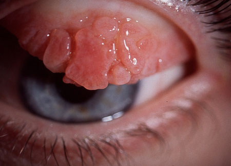
[Figure caption and citation for the preceding image starts]: BlepharitisPrivate collection of Dr Jonathan Smith and Dr Philip Severn; used with permission [Citation ends]. [Figure caption and citation for the preceding image starts]: Dry eye (stained with rose bengal)Private collection of Dr Jonathan Smith and Dr Philip Severn; used with permission [Citation ends].
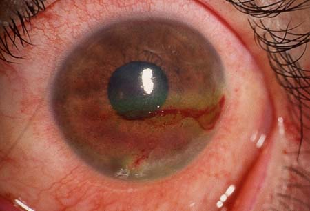
Examination for dry eye is particularly important before cataract and refractive surgery because its presence can adversely affect outcomes.
DED may be identified during routine comprehensive eye evaluations.[24]Chuck RS, Dunn SP, Flaxel CJ, et al. Comprehensive adult medical eye evaluation preferred practice pattern®. Ophthalmology. 2021 Jan;128(1):P1-29.
https://www.doi.org/10.1016/j.ophtha.2020.10.024
http://www.ncbi.nlm.nih.gov/pubmed/34933742?tool=bestpractice.com
Other tests
Schirmer's test assesses aqueous tear production and involves leaving Schirmer's paper strips in closed eyes for 5 minutes.[1]Hakim FE, Farooq AV. Dry eye disease: an update in 2022. JAMA. 2022 Feb 1;327(5):478-9.
http://www.ncbi.nlm.nih.gov/pubmed/35103781?tool=bestpractice.com
When performed without anaesthesia, Schirmer's test measures the total secretion (i.e., reflex and basic). When the test is performed with anaesthesia, it measures basic secretion.
Tear film osmometry measures tear film osmolarity. It is not widely available and is used in academic settings more often than in clinical practice.[3]Craig JP, Nichols KK, Akpek EK, et al. TFOS DEWS II definition and classification report. Ocul Surf. 2017 Jul;15(3):276-83.
https://www.sciencedirect.com/science/article/pii/S1542012417301192?via%3Dihub
http://www.ncbi.nlm.nih.gov/pubmed/28736335?tool=bestpractice.com
Other emerging tests include metalloproteinase-9 (MMP-9) detection, corneal topography, ocular surface interferometry, infrared thermography (assessing tear evaporation rate), in-vivo confocal microscopy, and ocular surface immune markers.[25]Lanza NL, Valenzuela F, Perez VL, et al. The matrix metalloproteinase 9 point-of-care test in dry eye. Ocul Surf. 2016 Apr;14(2):189-95.
http://www.ncbi.nlm.nih.gov/pubmed/26850527?tool=bestpractice.com
[26]Guven S. The repeatability of corneal topography measurements in severe dry eye disease. BMC Ophthalmol. 2022 Jul 15;22(1):306.
https://bmcophthalmol.biomedcentral.com/articles/10.1186/s12886-022-02534-4
http://www.ncbi.nlm.nih.gov/pubmed/35840937?tool=bestpractice.com
[27]Tangmonkongvoragul C, Chokesuwattanaskul S, Khankaeo C, et al. Prevalence of symptomatic dry eye disease with associated risk factors among medical students at Chiang Mai University due to increased screen time and stress during COVID-19 pandemic. PLoS One. 2022 Mar 23;17(3):e0265733.
https://journals.plos.org/plosone/article?id=10.1371/journal.pone.0265733
http://www.ncbi.nlm.nih.gov/pubmed/35320310?tool=bestpractice.com
[28]Tan LL, Sanjay S, Morgan PB. Screening for dry eye disease using infrared ocular thermography. Cont Lens Anterior Eye. 2016 Dec;39(6):442-9.
http://www.ncbi.nlm.nih.gov/pubmed/27568097?tool=bestpractice.com
[29]Gulias-Cañizo R, Rodríguez-Malagón ME, Botello-González L, et al. Applications of infrared thermography in ophthalmology. Life (Basel). 2023 Mar 8;13(3):723.
https://www.mdpi.com/2075-1729/13/3/723
http://www.ncbi.nlm.nih.gov/pubmed/36983878?tool=bestpractice.com
[30]Sim R, Yong K, Liu YC, et al. In vivo confocal microscopy in different types of dry eye and meibomian gland dysfunction. J Clin Med. 2022 Apr 22;11(9):2349.
https://www.mdpi.com/2077-0383/11/9/2349
http://www.ncbi.nlm.nih.gov/pubmed/35566475?tool=bestpractice.com
[31]Aluru SV, Agarwal S, Srinivasan B, et al. Lacrimal proline rich 4 (LPRR4) protein in the tear fluid is a potential biomarker of dry eye syndrome. PLoS One. 2012;7(12):e51979.
https://journals.plos.org/plosone/article?id=10.1371/journal.pone.0051979
http://www.ncbi.nlm.nih.gov/pubmed/23272196?tool=bestpractice.com
[32]Li Y, Zhang J, Liu X, et al. Identification of inflammatory markers as indicators for disease progression in primary Sjögren syndrome. Eur Cytokine Netw. 2024 Feb 1;35(1):1-12.
http://www.ncbi.nlm.nih.gov/pubmed/38909355?tool=bestpractice.com
[33]Aljohani S, Jazzar A. Tear cytokine levels in Sicca syndrome-related dry eye: a meta-analysis. Diagnostics (Basel). 2023 Jun 27;13(13):2184.
https://www.mdpi.com/2075-4418/13/13/2184
http://www.ncbi.nlm.nih.gov/pubmed/37443578?tool=bestpractice.com
[34]Roy NS, Wei Y, Yu Y, et al. Conjunctival HLA-DR expression and its association with symptoms and signs in the DREAM study. Transl Vis Sci Technol. 2019 Aug 21;8(4):31.
https://tvst.arvojournals.org/article.aspx?articleid=2748979
http://www.ncbi.nlm.nih.gov/pubmed/31489258?tool=bestpractice.com