Approach
In leprosy-endemic countries, diagnosis is made based on the clinical examination. However, laboratory tools are useful to confirm the diagnosis and to have an accurate classification.
History
Family members with leprosy, living in an endemic country, travel history, and contact with people with untreated leprosy, are important elements to consider. The average incubation period is thought to be between 3 and 5 years, although the minimum period has been reported as being as short as a few weeks, based on the occurrence of leprosy among young infants.[31] The maximum incubation period reported is ≥30 years, as observed among war veterans known to have been exposed for short periods in endemic areas but otherwise living in non-endemic areas.
Physical examination
Skin lesions can be single or multiple and can be hypopigmented or erythematous. A variety of skin lesions may be seen, but macules, papules, or nodules are common. The disease can also occur with multiple infiltrated patches or just diffuse skin infiltration. Sensory loss is a typical feature of leprosy; the skin lesion shows loss of sensation to pinprick and/or light touch.
Examination should include palpation of peripheral nerves. There may be tenderness, paraesthesias, or thickening of a nerve. Most commonly involved are the ulnar nerve, radial cutaneous nerve, median nerve, popliteal nerve, tibial nerve, and great auricular nerve. Numbness of extremities and loss of nerve function (as evidenced by claw hands, foot drop, facial paralysis) may also be present.
Pure neural leprosy is a rare presentation of leprosy and is found in 5% to 20% of patients.[32] Patients present with neuropathy without evidence of skin lesions.[33]
[Figure caption and citation for the preceding image starts]: Paucibacillary (PB) leprosy (borderline tuberculoid)WHO [Citation ends].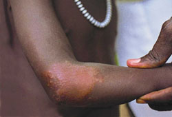 [Figure caption and citation for the preceding image starts]: Early multibacillary (MB) leprosyWHO [Citation ends].
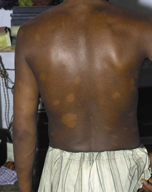 [Figure caption and citation for the preceding image starts]: Paucibacillary (PB) leprosy: tuberculoid (TT)WHO [Citation ends].
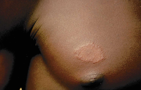 [Figure caption and citation for the preceding image starts]: Multibacillary (MB) leprosy nodulesWHO [Citation ends].
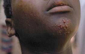
Investigations
Slit-skin smears using Wade-Fite stain can be taken when available. Rod-shaped, red-stained leprosy bacilli can be seen in multibacillary patients. Wherever possible, histopathology by a skin biopsy can be a valuable aid to differential diagnosis and for detailed classification of the disease. Nerve biopsy may be done in cases of pure neural leprosy.
Polymerase chain reaction may be used to detect Mycobacterium leprae DNA in tissue. It is useful to diagnose lepromatous leprosy (LL) but less sensitive to diagnose tuberculoid leprosy (TT) or borderline tuberculoid leprosy (BT).
Immunological reactions
Two types of reactions affect 30% to 50% of patients with leprosy: type 1 reaction (reversal reaction) and type 2 reaction (erythema nodosum leprosum). These reactions are often incorrectly viewed as complications of multi-drug therapy. Reactions are medical emergencies that can increase leprosy-related morbidity and so it is important to specifically recognise and treat reactions, to reduce the burden of disability in leprosy.
A third reaction, known as Lucio's phenomenon is relatively rare. Immunological reactions can occur any time, before, during, or after treatment.[5]
Type 1 reaction (reversal reaction)
Most commonly occurs in BT, mid-borderline leprosy (BB), borderline lepromatous (BL), LL
Existing skin lesions become erythematous and oedematous
Systemic symptoms are unusual
Neuritis: spontaneous nerve pain, tenderness, paraesthesias, and/or loss of nerve function (as evidenced by claw hand, foot drop, facial palsy) are commonly associated.
Type 2 reaction (erythema nodosum leprosum)
Most commonly occurs in BL and LL
Rapid appearance of crops of painful, erythematous subcutaneous nodules that can ulcerate
Fever, malaise, anorexia
Arthralgias
Orchitis, epididymitis, iritis
Neuritis.
Lucio's phenomenon
Occurs in diffuse non-nodular lepromatous leprosy (lepra bonita); has been associated with a different species called M lepromatosis[4]
Crops of haemorrhagic infarcts in the skin, plaques, which become necrotic and ulcerated
Atrophic scars left behind
Systemic symptoms are unusual.
Use of this content is subject to our disclaimer