Aetiology
Ascaris lumbricoides is the principal agent responsible for human ascariasis, although the pig nematode A suum has also produced human infection.[1][4][15][Figure caption and citation for the preceding image starts]: Photograph of 2 Ascaris lumbricoides nematodes; the larger one on the left is female and that on the right is male. Adult females can grow to >30 cm (12 in) in lengthPublic Health Image Library, CDC [Citation ends].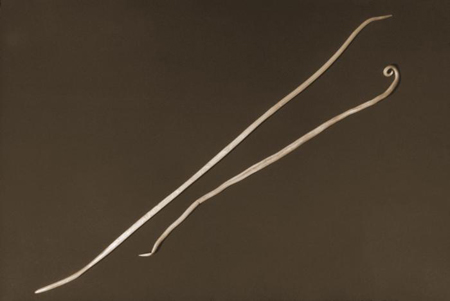
The life cycle begins with the production of mammillated eggs by adult females living in the distal small intestine of an infected human host. Once deposited in the soil, eggs become infectious within several weeks. Eggs are subsequently transmitted by ingestion, or possibly by inhalation of contaminated dust; larvae do not hatch in soil and do not invade the skin.
Inside their next human host, Ascaris larvae hatch in the jejunum, penetrate the intestinal wall, and migrate by way of hepatic venules to the right heart and pulmonary circulation. They subsequently break through into alveolar spaces, ascend the trachea, and are swallowed back into the intestine, where they undergo a final moult and develop into adults, which mate and spawn a new generation of eggs.
Occasionally, male-only adult infections occur, yielding no eggs in stool. Female-only infections produce infertile eggs, which never become infectious. Otherwise, under normal conditions, the time from ingestion of eggs to development of new eggs is 10 to 12 weeks. Adult worms can live for up to 18 months, and are then expelled.[1][Figure caption and citation for the preceding image starts]: A fertilised egg of the roundworm Ascaris lumbricoides at magnification x 400. Fertilised eggs are rounded, with a thick shell. Unfertilised eggs are elongated, are larger, have thinner shells, and are covered by a more visible mammillated layer, which is sometimes covered by protuberancesPublic Health Image Library, CDC [Citation ends].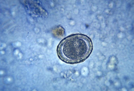
Pathophysiology
During larval migration through the lungs, eosinophilic pneumonitis, also known as Loeffler's syndrome, may develop in response to tissue destruction and released larval antigens.[1][13][16] This can manifest as asthma, with hypersecretion of mucus, bronchiolar inflammation, and serous exudate. Affected people may also have sputum containing eosinophils or Charcot-Leyden crystals (needle-shaped, pink, crystalline structures that result from the breakdown of eosinophils). In allergy-prone people, urticaria may also occur during larval migration.
Once larvae are ingested, they live in the small intestine and grow into adult worms measuring 15 cm to 40 cm (6 inches to 15.8 inches). Heavy worm burdens occasionally lead to intestinal and hepatobiliary obstruction, the main causes of morbidity and mortality due to ascariasis. The terminal ileum is the most common site of intestinal obstruction. Other complications result from the tendency of adult worms to migrate, an event often triggered by fever, medication, anaesthesia, or stress. Very rarely, aberrant ascarids have emerged through fistulas, or through the fallopian tubes, urinary bladder, lungs, or heart.
The most intense infections occur in children aged 5 to 15 years. Decreased exposure to eggs and acquired immunity account for lower worm burdens in adults.[17] The mechanism by which ascariasis stunts growth and development in children is not fully understood, but probably involves a combination of anorexia, protein and fat malabsorption, secondary lactose intolerance, and co-existing vitamin A deficiency. In one study, children with approximately 70 worms had a 72% impairment of dietary nitrogen absorption; two-thirds had moderate steatorrhoea.[18][Figure caption and citation for the preceding image starts]: Diagram depicting the various stages in the life cycle of the intestinal nematode Ascaris lumbricoidesPublic Health Image Library, CDC [Citation ends].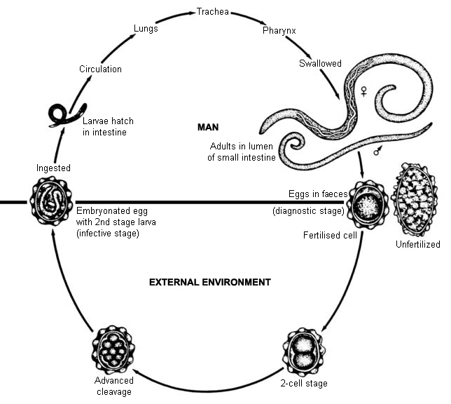
Use of this content is subject to our disclaimer