Images and videos
Images


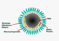

Rabies
Illustration of rabies virus in cross-section. Concentric layers: envelope membrane bilayer, M protein, and tightly coiled encased RNA
Centers for Disease Control and Prevention
See this image in context in the following section/s:
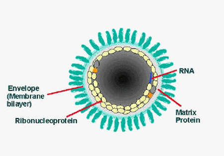
Rabies
Illustration of rabies virus in longitudinal section
Centers for Disease Control and Prevention
See this image in context in the following section/s:
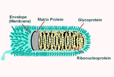
Rabies
A canine suspected of being rabid that had been exhibiting signs of restlessness and overall uncharacteristic aggressive behaviour
Centers for Disease Control and Prevention
See this image in context in the following section/s:
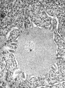
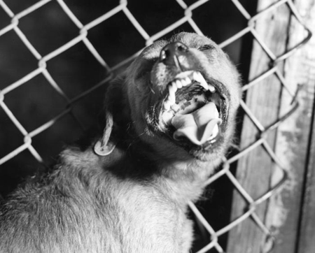
Rabies
This transmission electron micrograph reveals the presence of Lagos bat virus virions and an intracytoplasmic inclusion body in this tissue sample
Centers for Disease Control and Prevention; Dr Fred Murphy; Sylvia Whitfield
See this image in context in the following section/s:
Use of this content is subject to our disclaimer