Images and videos
Images
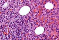

Hairy cell leukaemia
Sections of core biopsy demonstrate lymphocytes with obvious cytoplasm within the marrow interstitium, associated with dilatation of marrow sinuses and red blood cell collections (H&E 50x oil)
From the collection of Lynn Moscinski, MD
See this image in context in the following section/s:

Hairy cell leukaemia
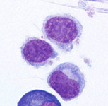
Cytospin prepared from bone marrow aspirate illustrates the typical cell cytology, with oval- to bean-shaped nuclei and moderate amounts of cytoplasm with irregular cytoplasmic borders (Wright Giemsa 100x oil)
From the collection of Lynn Moscinski, MD
See this image in context in the following section/s:
Use of this content is subject to our disclaimer