Aetiology
Eczema has a multifactorial aetiology, with a combination of genetic susceptibility and environmental factors contributing to disease development. Defects in the skin's barrier function and immune dysregulation following allergen exposure are key components in the development of this disease.
This finding has been linked to mutations in genes that are crucial for normal epidermal differentiation as well as to genes involved in immune-system regulation.[22][23] This genetic predisposition is supported by the increased incidence in family members, and by studies showing concordance rates of 77% in monozygotic twins and 15% in dizygotic twins.[21] Studies have estimated prevalence in siblings at 22% to 24%.[20]
Filaggrin gene mutations increase risk of eczema
As part of skin turnover, when granular layer cells differentiate into the stratum corneum (top layer of skin), profilaggrin is cleaved into filaggrin. Filaggrin breaks down to form a pool of amino acids that are essential for an effective skin barrier. Loss‐of‐function mutations in the filaggrin gene (located in the epidermal differentiation complex on chromosome 1q21.3 I) predispose to breaks in the epidermal barrier, allowing increased exposure and sensitisation to antigens, and to increased risk for eczema.[23][24][25][26]
Links have also been identified between eczema and areas of the genome that are known to encode cytokines and receptors involved in the Th2-mediated immune response that predominates in eczema.[21]
Environmental factors
Acquired insults to the mechanical skin barrier may trigger or worsen the disease state.[27][28] The role of environmental factors in the development of eczema is supported by the increased rate of disease seen in urban areas, smaller families, and higher socioeconomic classes.[17] In one study, the prevalence of eczema in Jamaican children living in London was twice as high as that of Jamaican children living in Jamaica.[10] In the US the rate of prevalence and persistence of eczema is higher for children who are female or black and from an urban environment.[18]
Environmental factors that can trigger or exacerbate eczema include:[27][28][29][30][31][32]
Irritants such as soaps and detergents (including shampoos, bubble baths)
Skin infections (Staphylococcus aureus infection is a common cause of eczema exacerbation)
Contact allergens
Food allergens (cows' milk, egg, nuts)
Inhalant allergens (dust mite)
Vitamin E deficiency
Hard domestic water supply (high calcium carbonate).
There is evidence to suggest that being born during the autumn and winter months in the northern hemisphere is significantly associated with the development of eczema. Further studies are required to understand the effect of seasonal change.[33]
Pathophysiology
Impairment of the skin's barrier function leads to an increased sensitisation to cutaneous antigens and is a major factor in the pathophysiology.[7] Mutations in the filaggrin gene, which encodes a protein necessary for terminal differentiation of epidermis, result in impaired barrier function of the epidermis and increased exposure and sensitisation to cutaneous antigens.[23]
In the acute phase of eczema, the immune response following sensitisation is predominantly Th2 mediated, with over-expression of IL-4, IL-5, and IL-13. These interleukins lead to an increased production of IgE and peripheral eosinophilia.[1][34]
Susceptibility has been linked to polymorphisms in the gene encoding a subunit of the IgE receptor as well as to a region on chromosome 5q31-33 that includes genes for cytokines expressed by Th2 cells.[21]
Persistent inflammation and scratching can eventually lead to chronic eczema, with thick, lichenified skin. Lesions demonstrate a different complement of immune cells and cytokines, with a predominant Th1 response, and increased levels of IL-12.[26] Higher expression of Th17 and IL-17 have also been associated with chronic eczema.[35]
Classification
Classification by age: infantile, childhood, and adult phases[1]
Although there is not a standardised classification system for eczema, the disease is commonly divided into three phases: infantile, childhood, and adult.[1]
While the phases are typical of eczema, it is important to note that patients will not necessarily progress through each phase. For example, symptoms may resolve after childhood or may initially present later in life. There are no distinct cut-off ages for each phase.
The infantile phase:[1]
Typically lasts from shortly after birth to 2 years of age
Often begins with dermatitis on the cheeks, forehead, and scalp with significant involvement of extensor surfaces of the limbs
Commonly a prominent vesicular component with oedema, weeping, and crusting
Facial eczema may be worse while infants are teething and with trials of new foods
Extensor surface involvement may be related to the onset of crawling.
Lasts from 2 years of age until puberty; children with already persistent disease, later onset, and/or more severe disease have increased persistence
Affected areas are typically less vesicular, with papules and plaques becoming more lichenified due to constant scratching
Children typically have involvement of the flexural skin, with predominance in the antecubital and popliteal fossae, wrists, hands, ankles, and feet
When facial involvement is present, it is typically confined to peri-oral and peri-orbital skin.[Figure caption and citation for the preceding image starts]: Acute eczema in the antecubital fossa of a 9-year-old girlFrom the personal collection of A. Hebert, MD; used with permission [Citation ends].

The adult phase:[1]
Extends from puberty through adulthood
Thickened, dry skin and lichenified plaques are typical; flexural skin, the upper back and arms, as well as the dorsal surfaces of the hands and feet, are often affected
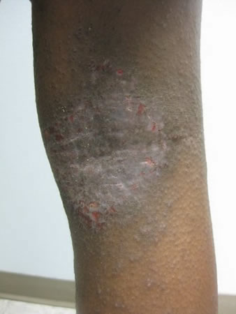
Dyshidrotic changes may be present on the palms and soles.[Figure caption and citation for the preceding image starts]: Lichenification of the popliteal fossa in a child with eczemaFrom the personal collection of A. Hebert, MD; used with permission [Citation ends].
 [Figure caption and citation for the preceding image starts]: Chronic eczema affecting the palm of a 64-year-old manFrom the personal collection of A. Hebert, MD; used with permission [Citation ends].
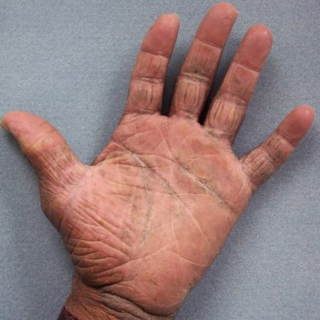
Eczema Area and Severity Index[3]
In secondary care, eczema severity is increasingly measured using the Eczema Area and Severity Index (EASI). EASI is a clinical tool scored by assessing the erythema, excoriations, induration, and lichenification of the skin. DermNet NZ: EASI score Opens in new window
UK and European guidelines require that outcomes of patients receiving dupilumab are reported using EASI (which has influenced its uptake as a clinical disease severity tool for eczema in the UK and Europe).[4][5]
Scoring of Atopic Dermatitis[3]
A composite score developed by the European Task Force on Atopic Dermatitis. Scoring of Atopic Dermatitis (SCORAD) values below 25 are considered to be mild eczema; SCORAD above 50 is regarded as severe. DermNet NZ: SCORAD Opens in new window
Use of this content is subject to our disclaimer