Images and videos
Images


Assessment of lymphadenopathy
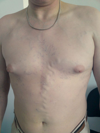
Prominent collateral vein on the anterior chest and abdominal wall in a patient with superior vena cava syndrome
From the collection of E Kempny
See this image in context in the following section/s:

Assessment of lymphadenopathy
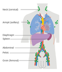
Diagram showing the lymph node groups most commonly affected by Hodgkin’s lymphoma
Cancer Research UK
See this image in context in the following section/s:
Assessment of lymphadenopathy
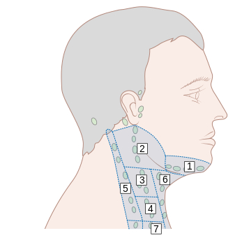
Lymph node groups in the head and neck; the numbers refer to the anatomical levels of the lymph nodes
Cancer Research UK
See this image in context in the following section/s:
Assessment of lymphadenopathy
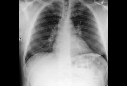
Bilateral hilar adenopathy
From the collection of Dr M.P. Muthiah; used with permission
See this image in context in the following section/s:
Use of this content is subject to our disclaimer