Images and videos
Images


Non-sustained ventricular tachycardias
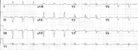
ECG showing an abnormal Q wave in V4 and ST elevation in V1-V6 in a patient with a large anterior wall myocardial infarction
Kusumoto FM. ECG interpretation. In: Pathophysiology to clinical application. New York, NY: Springer; 2009; used with permission
See this image in context in the following section/s:

Non-sustained ventricular tachycardias
Non-sustained ventricular tachycardia
From the collection of Dr F. Kusumoto; used with permission
See this image in context in the following section/s:

Non-sustained ventricular tachycardias
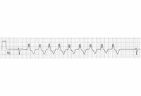
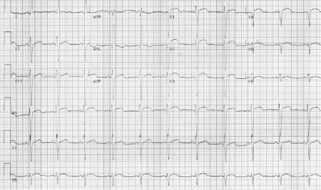
ECG from a patient with Brugada's syndrome, showing terminal positive R-wave and ST-segment elevation in lead V1
Kusumoto FM. ECG interpretation. In: Pathophysiology to clinical application. New York, NY: Springer; 2009; used with permission
See this image in context in the following section/s:
Non-sustained ventricular tachycardias
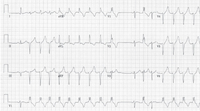
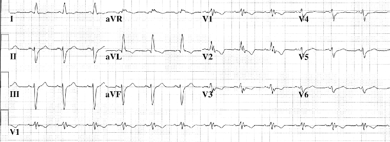
Initiation of wide QRS rhythm is not preceded by a P-wave, suggesting non-sustained ventricular tachycardia
Kusumoto FM. ECG interpretation. In: Pathophysiology to clinical application. New York, NY: Springer; 2009; used with permission
See this image in context in the following section/s:

Non-sustained ventricular tachycardias
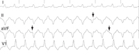
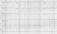
ECG showing AV dissociation. Deflections due to P waves (arrows) are not associated with QRS complexes
Kusumoto FM. ECG interpretation. In: Pathophysiology to clinical application. New York, NY: Springer; 2009; used with permission
See this image in context in the following section/s:
Non-sustained ventricular tachycardias
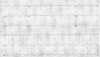
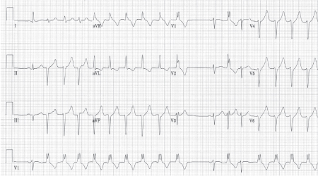
ECG from a patient with arrhythmogenic right ventricular cardiomyopathy
Kusumoto FM. ECG interpretation. In: Pathophysiology to clinical application. New York, NY: Springer; 2009; used with permission
See this image in context in the following section/s:
Non-sustained ventricular tachycardias
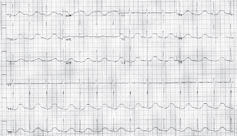
ECG from a patient with long QT syndrome
Kusumoto FM. ECG interpretation. In: Pathophysiology to clinical application. New York, NY: Springer; 2009; used with permission
See this image in context in the following section/s:
Non-sustained ventricular tachycardias
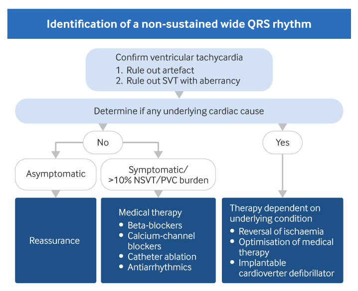
Evaluation of non-sustained wide QRS tachycardia
Created by BMJ Knowledge Centre.
See this image in context in the following section/s:
Use of this content is subject to our disclaimer