Approach
Most MCL injuries are treated conservatively. Rest, ice, compression, and elevation (RICE), protective ambulation (ambulation with crutches or other assistive device), and physiotherapy are commonly employed treatment measures.
For treatment purposes, the American Medical Association classification of MCL injuries is used. MCL injuries are classified based on the amount of medial joint opening when a valgus load is applied at 20 to 30 degrees of knee flexion:[2]
Grade I: 0 to 5 mm of opening
Grade II: 5 to 10 mm of opening
Grade III: >10 mm of opening
Acute treatment
Initially, all suspected knee sprains should be treated with the RICE protocol: rest, ice, compression of the knee with an elastic bandage, and elevation of the leg. Non-steroidal anti-inflammatory drugs can be given to further reduce swelling and provide pain relief. If the injured knee is unstable or exceptionally painful, a hinged knee brace or an immobilising knee brace and/or crutches should be used. Grade I injuries do not usually require protective ambulation but grade II and III injuries usually do. Grade III MCL injuries should be immobilised with a hinged knee brace set at 30 degrees flexion to minimise the distance between the two ends of the torn ligament.[29] A hinged brace that allows full flexion but minimises full extension is recommended for minimising strain on the MCL and protecting against further injury. It is critical that the brace has sufficient rigidity to stabilise medial and lateral movement. The brace should be worn for 4 to 6 weeks.[Figure caption and citation for the preceding image starts]: Hinged knee braceFrom the collection of Sanjeev Bhatia, MD; used with permission [Citation ends].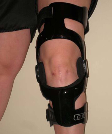
Physiotherapy
After swelling subsides and the patient can ambulate more easily, the patient should undertake a physiotherapy regimen aimed at improving quadriceps and hamstring strength. Most MCL injuries, even grade III, heal well non-operatively. Physiotherapy restores range of movement and improves muscle strength, resulting in decreased pain and increased knee function. Therapy begins with low-impact exercises, gradually progressing to more sport/activity-specific exercises. When sport-specific exercises, particularly those involving cutting and pivoting, can be performed without discomfort, patients are allowed to return to competitive play. Postoperative physiotherapy is an important part of the rehabilitation process, which all patients should receive.
Surgery
MCL injuries rarely require surgical reconstruction. The exceptions are chronic MCL injuries (≥3 months' duration with high-grade laxity) that have failed non-operative treatment[18][30][31][4] and certain multiple-ligament knee injuries. Additionally, tibial-sided grade III MCL injuries are more prone to persistent laxity even after conservative management. These patients frequently have valgus instability even with activities of daily living.[30]
Surgery for isolated grade III MCL injury is sometimes indicated.[32] Acute grade III MCL injuries may warrant operative intervention if there is also a large bony avulsion, tibial plateau fracture, intra-articular entrapment of the end of a ligament, or anteromedial instability (positive anterior drawer test). [Figure caption and citation for the preceding image starts]: The anterior drawer testFrom the collection of Sanjeev Bhatia, MD; used with permission [Citation ends].
If undertaken, surgical repair is usually carried out 7 to 10 days after injury.[17][18][31] In many cases some mild degree of persistent instability remains even after successful reconstruction. Specific complications are unusual but include decreased range of motion (ROM) (if the MCL graft is placed in a non-anatomical position) and saphenous nerve injury.
In multi-ligament knee injuries, MCL reconstruction is usually warranted, as the healing process may be compromised due to the functional loss of other ligaments[33] and so predisposing to chronic MCL insufficiency.[30]
In combined MCL and anterior cruciate ligament (ACL) injuries, ACL reconstruction is generally recommended after a period of rehabilitation to allow the MCL to heal.[34] Surgery is performed after achieving full ROM, adequate strength, and resolution of knee effusion.[7][18][35] This usually occurs 4 to 6 weeks after injury, when the ACL can then be reconstructed with a patellar tendon graft or hamstring tendon graft. An autograft or allograft tendon can be used with excellent results.
If valgus instability persists after ACL reconstruction, the patient should undergo surgical MCL reconstruction.[36][37] MCL reconstruction may also be warranted if there is a large bony avulsion, tibial plateau fracture, intra-articular entrapment of the end of a ligament, or anteromedial instability (positive anterior drawer test).
In combined MCL and non-anterior ligament injuries, MCL repair is usually performed 7 to 10 days after injury. Surgical reconstruction or repair of the other injured ligament (posterior cruciate ligament, meniscus, lateral collateral ligament) is usually warranted within 3 weeks of injury. Compared with non-surgical management or a delay in surgery, early operative treatment of the multi-ligament-injured knee yields improved functional and clinical outcomes.[38][39] In one meta-analysis, a staged approach to reconstruction yielded the best outcomes with regard to subjective scores and range of motion.[32] Reconstruction of the posterolateral corner is preferred over repair as it results in decreased revision rates.[38]
Both chronic MCL injuries and multiple knee ligament injuries should be referred to an orthopaedic specialist. Isolated grade III MCL injuries in high-demand athletes and labourers should also be referred for consultation.
Use of this content is subject to our disclaimer