Approach
A typical diagnosis starts with careful questioning to determine a patient's symptoms (e.g., gastrointestinal symptoms, infertility, haematuria, haematospermia, or vaginal/vulvar lesions) and whether the patient has lived in or visited an area endemic for schistosomiasis. Thereafter, specific physical examinations and laboratory tests allow for diagnosis and/or referral.
History
Travel and residence history is the most important element in defining risk of exposure to infection.[36] Schistosomiasis is found in Africa, the Middle East, the Far East, and in parts of South America and the Caribbean.[14] A history of activities involving freshwater exposure in endemic areas is also important to assess risk. For travellers, precaution is most important. For the population in endemic areas, an integrated approach including health education is necessary.
The time interval since exposure and the duration of symptoms can help differentiate between skin infection (cercarial dermatitis), acute disease (Katayama syndrome), and chronic disease. Acute schistosomiasis occurs several weeks after exposure. Its symptoms are non-specific, including fever, fatigue, cough, abdominal pain, and diarrhoea. Chronic intestinal schistosomiasis may present with symptoms of intermittent abdominal pain, diarrhoea, and bloody stools. Urinary schistosomiasis may present with dysuria, lower abdominal pain, infertility, or a history of haematuria, haematospermia, or vaginal or vulvar lesions. Progressive obliteration of pulmonary arterioles and capillaries occurs and leads to secondary fibrotic changes in the lung parenchyma, often over decades. This may manifest as progressive dyspnoea.
Physical examination
A papular rash may be found on skin exposed to contaminated water hours after exposure (swimmer's itch).[37][Figure caption and citation for the preceding image starts]: Mild cercarial dermatitis (swimmer's itch)CDC; used with permission [Citation ends].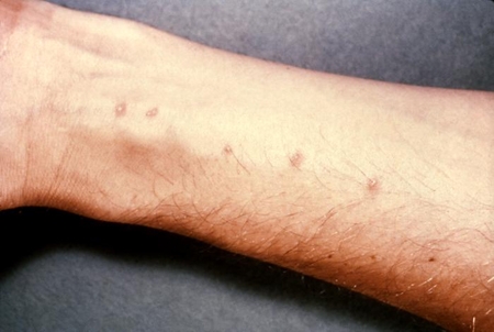
Fever, cough, abdominal tenderness, or hepatosplenomegaly may be observed in acute schistosomiasis 1 to 3 months after exposure. Examination findings are often non-specific at this stage of infection.[19][38][39]
Growth retardation, weight loss, and signs of anaemia may be detected in children with a chronic infection.[22][40][41] Other findings in patients with the chronic intestinal form of schistosomiasis include signs of portal hypertension including ascites, pedal oedema, and hepatosplenomegaly.[1] In urinary schistosomiasis, hydronephrosis might be detected as an abdominal mass on examination, and hepatosplenomegaly may also be present. Abdominal or pelvic tenderness can often be detected. In women, a pelvic examination may show ulceration of the vulvar skin or vaginal or cervical surfaces.[42] For men, a rectal examination may reveal prostatic mass, tenderness, or enlargement.[43]
Laboratory examination
The most specific test for infection is detection of Schistosoma eggs in urine, faeces, or tissue biopsy.[44] Although dead eggs can be passed for some time after infection is gone, parasitological detection of eggs should always be treated with praziquantel to eliminate a potential active infection. A negative egg screen does not exclude active infection. These parasitological tests can be negative during acute infection and during a low-intensity chronic infection.[45]
Positive serology for anti-Schistosoma antibodies is sensitive and specific for exposure to infection but cannot discriminate between current active and past infection, or determine parasite burden.[46] Also, the serological tests may take 3 months to become positive, so individuals with clinical manifestations of acute schistosomiasis commonly have negative serological tests. An untreated person with a positive serology and a <5-year interval since travel exposure should be considered for presumptive praziquantel therapy of chronic infection.
Other supportive (but not pathognomonic) diagnostic tests include abnormalities found on stool guaiac test for occult gastrointestinal bleeding; an FBC detecting anaemia or eosinophilia; urinalysis for haematuria, proteinuria, pyuria, and eosinophiluria; and an abnormal renal function panel or liver function test. Where the index of suspicion for schistosomiasis is high and standard parasitological testing is negative, these abnormalities should prompt serological testing, imaging studies, or tissue biopsy to look for other evidence of active Schistosoma infection.
Blood cultures and malaria thick and thin smears are normally performed in recently returned travellers from endemic areas with suspected acute schistosomiasis cases, to rule out other causes of fever. These results should be negative if schistosomiasis is the only source of fever.
A number of tests designed to detect schistosome DNA or RNA have been developed to complement parasitological and serological diagnosis with high reported accuracy. The availability of these tests varies depending on location. Tests can be run on serum, faeces, CSF, or urine.[47]
Urinary circulating anodic antigen (CAA) is a point of care test that is used for screening for urinary schistosomiasis. It is used in endemic areas where there is not adequate access to microscopy. This test has good sensitivity; however, specificity is controversial as a good number of individuals have positive tests in the absence of positive microscopy. This may be because it is more sensitive than microscopy.[47][48]
[Figure caption and citation for the preceding image starts]: Micrograph depicting an egg from a Schistosoma haematobium trematode parasite; magnified 500xCDC; used with permission [Citation ends].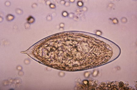
Radiological examination
Ultrasound examination, CT, or MRI of affected organs (bladder and kidneys or portal veins and liver) can often show characteristic changes that are helpful in the diagnosis and follow-up of treatment. MRI of the brain or spinal column is indicated for neurological symptoms.[49] Chest x-ray and chest CT are indicated for evaluation of suspected pulmonary hypertension in patients with respiratory symptoms.[50]
Indications for referral
Ulceration of the vagina or cervix should be biopsied to exclude cancer.[42] Infertility requires more extensive anatomical and endocrine evaluation for other causes, and gynaecological consultation is advised.[51]
Neuroschistosomiasis presents with seizures or focal neurological symptoms related to the site of inflammation (i.e., brain, spinal cord, leptomeninges, or peripheral nerve roots).[28][49][52][53] Early spinal cord MRI and neurological and neurosurgical consultation is recommended for management in these cases.
Advanced portal hypertension from intestinal schistosomiasis may present with ascites and severe upper gastrointestinal bleeding from oesophageal varices.[54] Consultation with a gastroenterologist and/or invasive radiologist is recommended for management of these cases.
Late-stage infection may also present with pulmonary symptoms and signs of cor pulmonale.[50] In these cases, pulmonary or cardiology consultation is recommended to exclude other aetiologies.
Advanced urinary schistosomiasis may present with obstructive uropathy and signs of renal failure, or with evidence of bladder cancer.[55] Here, urology and nephrology consultation is recommended for disease management.
Use of this content is subject to our disclaimer