Summary
Definition
History and exam
Key diagnostic factors
- presence of risk factors
- numbness
- weakness
- pain
- sicca symptoms
- parotid gland enlargement
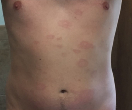
- rash, ulcerations, or pigment changes
- wheeze, cough, other pulmonary signs
- fever, night sweats, weight loss, and malaise
Other diagnostic factors
- predisposing conditions causing vasculitis, inflammation, or other nerve damage
Risk factors
- age over 50 years
- sarcoidosis
- hepatitis C
- cryoglobulinaemia
- hepatitis B
- connective tissue disease
- livedo reticularis
- primary vasculitis
- medications
- HIV infection
- non-HIV, non-hepatitis infections
- severe acute respiratory syndrome coronavirus-2 (SARS-CoV-2) infection
- recreational drug use
- genetic predisposition
Diagnostic investigations
1st investigations to order
- electromyogram (EMG)
- FBC with differential
- erythrocyte sedimentation rate (ESR)
- C-reactive protein (CRP)
- serum creatinine
- serum glucose
- cryoglobulins
- serum complement
- hepatitis B surface antigen
- hepatitis C antibodies or RNA
- anti-HIV antigens or HIV RNA
- Lyme disease antibodies
- cytoplasmic and perinuclear antineutrophil cytoplasmic antibodies (c-ANCA and p-ANCA)
- rheumatoid factor
- antinuclear antibodies (ANA)
- antidouble-stranded (ds) DNA
- anti-Sjogren syndrome-related antigen A (SSA) or -SSB antibodies
- serum angiotensin-converting enzyme
- protein electrophoresis and immunofixation
- chest x-ray
- urinalysis
- muscle and nerve biopsy
Investigations to consider
- anti-Smith (anti-Sm) antibodies
- anti-topoisomerase I (anti-Scl 70) and anticentromere (ACA) antibodies
- skin biopsy
- lip biopsy
- anti-Hu antibodies
- cerebrospinal fluid (CSF) analyses
- CT of chest, abdomen/pelvis
- positron emission tomography (PET) scan of chest, abdomen, or pelvis
- conventional angiography
- magnetic resonance angiography
Treatment algorithm
Contributors
Authors
Ashok Verma, MD, DM, MBA, FAAN, FANA
Professor of Neurology
Director, Neuromuscular Medicine Fellowship Training Program
University of Miami Miller School of Medicine
Don Soffer Clinical Research Center
Miami
FL
Disclosures
AV declares that he has no competing interests.
Acknowledgements
Professor Ashok Verma would like to gratefully acknowledge Dr Kevin Scott, Dr Milind Kothari, and Dr Jenice Robinson, the previous contributors to this topic.
Disclosures
JR, KS, and MK declare that they have no competing interests.
Peer reviewers
John J. Kelly, MD
Professor and Chairman
Department of Neurology
The George Washington University Medical Center
Washington
DC
Disclosures
JJK declares that he has no competing interests.
Cory Toth, BSc, MD, FRCP(C)
Assistant Professor of Neurosciences
Hotchkiss Brain Institute
University of Calgary
Alberta
Canada
Disclosures
CT declares that he has no competing interests.
Jeremy Bland, FRCP
Consultant Neurophysiologist
East Kent Hospitals NHS Trust
Canterbury
Kings College Hospital NHS Trust
London
UK
Disclosures
JB declares that he has no competing interests.
Use of this content is subject to our disclaimer