The initial assessment of suspected asthma is focused on the presence of key features in the history, clinical examination, and medical records, together with careful consideration of risk factors and alternative diagnoses.[1]Global Initiative for Asthma. 2024 Global strategy for asthma management and prevention. May 2024 [internet publication].
https://ginasthma.org/wp-content/uploads/2024/05/GINA-2024-Strategy-Report-24_05_22_WMS.pdf
[98]Moral L, Vizmanos G, Torres-Borrego J, et al. Asthma diagnosis in infants and preschool children: a systematic review of clinical guidelines. Allergol Immunopathol (Madr). 2019 Mar-Apr;47(2):107-21.
http://www.ncbi.nlm.nih.gov/pubmed/30193886?tool=bestpractice.com
A probability-based approach plus a therapeutic trial is recommended for children 5 years and younger because most will be unable to perform lung function tests reliably. Children 6 years and older should have their asthma diagnosis confirmed with a test of variable expiratory airflow limitation.[1]Global Initiative for Asthma. 2024 Global strategy for asthma management and prevention. May 2024 [internet publication].
https://ginasthma.org/wp-content/uploads/2024/05/GINA-2024-Strategy-Report-24_05_22_WMS.pdf
[99]Gupta S, Thériault G. Do not diagnose or routinely treat asthma or chronic obstructive pulmonary disease without pulmonary function testing. BMJ. 2023 Mar 20;380:e072834.
http://www.ncbi.nlm.nih.gov/pubmed/36940980?tool=bestpractice.com
[100]National Institute for Health and Care Excellence. Asthma: diagnosis, monitoring and chronic asthma management. Mar 2021 [internet publication].
https://www.nice.org.uk/guidance/ng80
Although the peak expiratory flow (PEF) is less reliable than spirometry, its use is recommended where diagnosis would otherwise rely on symptoms only.[1]Global Initiative for Asthma. 2024 Global strategy for asthma management and prevention. May 2024 [internet publication].
https://ginasthma.org/wp-content/uploads/2024/05/GINA-2024-Strategy-Report-24_05_22_WMS.pdf
Definitive diagnosis requires a history of reversible airway obstruction and symptom improvement with inhaled bronchodilators or corticosteroids. See Diagnostic criteria for more information.The basis of an asthma diagnosis or treatment decision should be clearly documented.[1]Global Initiative for Asthma. 2024 Global strategy for asthma management and prevention. May 2024 [internet publication].
https://ginasthma.org/wp-content/uploads/2024/05/GINA-2024-Strategy-Report-24_05_22_WMS.pdf
[8]British Thoracic Society. British guideline on the management of asthma. Jul 2019 [internet publication].
https://www.brit-thoracic.org.uk/quality-improvement/guidelines/asthma
See Acute asthma exacerbation in children for more information about the diagnosis of acute exacerbations.
History
Recurrent symptoms of wheezing, cough (worse at night or early morning), and shortness of breath in response to recognised triggers (e.g., temperature change, viral infection, exercise, and emotion) are characteristic of asthma. Parental perception should be checked because various respiratory noises may be incorrectly labelled as wheezing (where possible, wheeze should be confirmed by a healthcare professional).[101]Cane RS, Ranganathan SC, McKenzie SA. What do parents of wheezy children understand by "wheeze"? Arch Dis Child. 2000 Apr;82(4):327-32.
https://adc.bmj.com/content/82/4/327.long
http://www.ncbi.nlm.nih.gov/pubmed/10735844?tool=bestpractice.com
The diagnosis is supported by features of atopic disease, such as eczema, atopic dermatitis, and allergic rhinitis in the child or first-degree family members.
Features associated with asthma include
Episodic symptoms (wheeze, breathlessness, chest tightness, and cough occurring episodically with periods of no or minimal symptoms)
Wheeze confirmed by a healthcare professional
Diurnal variability (worse at night or early morning)
A history of atopy
Recurrent events over time
Cough is often misdiagnosed as asthma in children, and requires a careful review of the history and the exclusion of alternative causes (see Differentials).[102]Lai K, Satia I, Song WJ, et al. Cough and cough hypersensitivity as treatable traits of asthma. Lancet Respir Med. 2023 Jul;11(7):650-62.
http://www.ncbi.nlm.nih.gov/pubmed/37336227?tool=bestpractice.com
Many children younger than 5 years present with recurrent wheezing due to frequent upper respiratory tract infections (URTIs).[1]Global Initiative for Asthma. 2024 Global strategy for asthma management and prevention. May 2024 [internet publication].
https://ginasthma.org/wp-content/uploads/2024/05/GINA-2024-Strategy-Report-24_05_22_WMS.pdf
Wheeze is a heterogeneous phenotype in young children, and many non-atopic children who experience recurrent episodes of viral-induced wheezing will not require a regular ICS or go on to develop chronic atopic asthma.
Physical examination
Most children have no signs when they do not have an exacerbation. Depending on the symptom pattern, examination may uncover widespread polyphonic wheeze audible on chest auscultation and respiratory distress (e.g., tachypnoea, recessions or retractions, and accessory muscle use). In poorly controlled persistent asthma, hyperinflation (reflecting gas trapping) and chest wall deformity (Harrison's sulci) may be present. Features of atopic disease may be evident on examination.
Probability-based approach to diagnosis
The greater the variations in expiratory lung function, and the more often excess variation is observed, the more confident a clinician can be with the diagnosis of childhood asthma.[1]Global Initiative for Asthma. 2024 Global strategy for asthma management and prevention. May 2024 [internet publication].
https://ginasthma.org/wp-content/uploads/2024/05/GINA-2024-Strategy-Report-24_05_22_WMS.pdf
However, it is important to ensure that apparent variability does not reflect variations in technique over time, because both spirometry and PEF measures are effort-dependent. The following probability-based approach to diagnosis is proposed.[8]British Thoracic Society. British guideline on the management of asthma. Jul 2019 [internet publication].
https://www.brit-thoracic.org.uk/quality-improvement/guidelines/asthma
[103]National Heart, Lung, and Blood Institute; National Asthma Education and Prevention Program. Guidelines for the diagnosis and management of asthma. Aug 2007 [internet publication].
http://www.nhlbi.nih.gov/health-pro/guidelines/current/asthma-guidelines/full-report.htm
High probability of asthma: start with a therapeutic trial and reserve further testing for those with a poor response.
Intermediate probability of asthma (can perform spirometry and has evidence of airway obstruction): offer a reversibility test and/or time-limited therapeutic trial.
If there is reversibility, or if treatment is beneficial, treat as asthma.
If there is no significant reversibility, and/or a therapeutic trial is not beneficial, consider tests for alternative conditions.
Intermediate probability of asthma (can perform spirometry and with no evidence of airway obstruction): consider testing for atopic status, bronchodilator reversibility, and, if possible, bronchial hyper-responsiveness using methacholine or exercise.
Intermediate probability of asthma (unable to perform spirometry): consider testing for atopic status and offering a time-limited therapeutic trial.
If beneficial, treat as asthma.
If not beneficial, stop treatment and consider an alternative diagnosis and/or specialist referral.
Low probability of asthma: consider more detailed investigation (particularly for other diagnoses) and specialist referral.
The basis on which the diagnosis is suspected should be recorded. Using a structured questionnaire can help standardise this approach, but reliance on asthma prediction tools and screening aids is not recommended because they show wide variation in performance when assessing future risk.[104]Chang TS, Lemanske RF Jr, Guilbert TW, et al. Evaluation of the modified asthma predictive index in high-risk preschool children. J Allergy Clin Immunol Pract. 2013 Mar;1(2):152-6.
https://www.ncbi.nlm.nih.gov/pmc/articles/PMC3811153
http://www.ncbi.nlm.nih.gov/pubmed/24187656?tool=bestpractice.com
[105]Colicino S, Munblit D, Minelli C, et al. Validation of childhood asthma predictive tools: A systematic review. Clin Exp Allergy. 2019 Apr;49(4):410-18.
http://www.ncbi.nlm.nih.gov/pubmed/30657220?tool=bestpractice.com
[106]Kothalawala DM, Kadalayil L, Weiss VBN, et al. Prediction models for childhood asthma: a systematic review. Pediatr Allergy Immunol. 2020 Aug;31(6):616-27.
http://www.ncbi.nlm.nih.gov/pubmed/32181536?tool=bestpractice.com
[107]Daines L, McLean S, Buelo A, et al. Systematic review of clinical prediction models to support the diagnosis of asthma in primary care. NPJ Prim Care Respir Med. 2019 May 9;29(1):19.
http://www.ncbi.nlm.nih.gov/pubmed/31073125?tool=bestpractice.com
The Childhood Asthma Risk Tool (CHART) has been proposed; its performance in clinical care remains unclear.[108]Reyna ME, Dai R, Tran MM, et al. Development of a symptom-based tool for screening of children at high risk of preschool asthma. JAMA Netw Open. 2022 Oct 3;5(10):e2234714.
http://www.ncbi.nlm.nih.gov/pubmed/36201211?tool=bestpractice.com
Response to medication
Response to a therapeutic trial of inhaled beta-2 agonist or corticosteroid, given as either a short course of oral corticosteroid (e.g., 1-2 mg/kg/day for 3 days) or a longer trial of low-dose inhaled corticosteroid (e.g., for 4-6 weeks), is suggestive of asthma.[1]Global Initiative for Asthma. 2024 Global strategy for asthma management and prevention. May 2024 [internet publication].
https://ginasthma.org/wp-content/uploads/2024/05/GINA-2024-Strategy-Report-24_05_22_WMS.pdf
[8]British Thoracic Society. British guideline on the management of asthma. Jul 2019 [internet publication].
https://www.brit-thoracic.org.uk/quality-improvement/guidelines/asthma
Any therapeutic trial should be time-limited:
Consider a diagnosis of asthma if there is clinical improvement (based on symptom control and exacerbation rate) during treatment and deterioration when treatment is stopped.
Consider alternative diagnoses if there is a lack of response; cough and wheeze often have different causes in children than in adults, necessitating care to ensure proper investigation.[102]Lai K, Satia I, Song WJ, et al. Cough and cough hypersensitivity as treatable traits of asthma. Lancet Respir Med. 2023 Jul;11(7):650-62.
http://www.ncbi.nlm.nih.gov/pubmed/37336227?tool=bestpractice.com
This approach is also suitable for children with a non-specific cough and risk factors for asthma.[102]Lai K, Satia I, Song WJ, et al. Cough and cough hypersensitivity as treatable traits of asthma. Lancet Respir Med. 2023 Jul;11(7):650-62.
http://www.ncbi.nlm.nih.gov/pubmed/37336227?tool=bestpractice.com
[109]Morice AH, Millqvist E, Bieksiene K, et al. ERS guidelines on the diagnosis and treatment of chronic cough in adults and children. Eur Respir J. 2020 Jan;55(1):1901136.
http://www.ncbi.nlm.nih.gov/pubmed/31515408?tool=bestpractice.com
[110]Chang AB, Oppenheimer JJ, Irwin RS, et al. Managing chronic cough as a symptom in children and management algorithms: CHEST guideline and expert panel report. Chest. 2020 Jul;158(1):303-29.
http://www.ncbi.nlm.nih.gov/pubmed/32179109?tool=bestpractice.com
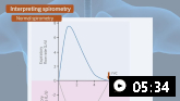
Spirometry
Spirometry is the preferred method for assessing variability in expiratory lung function. It is performed in children with suspected asthma who are able to give repeatable and reproducible results, but this is highly dependent on local service availability and the child's level of cooperation (typically from age 5 years).[1]Global Initiative for Asthma. 2024 Global strategy for asthma management and prevention. May 2024 [internet publication].
https://ginasthma.org/wp-content/uploads/2024/05/GINA-2024-Strategy-Report-24_05_22_WMS.pdf
[111]Gaillard EA, Kuehni CE, Turner S, et al. European Respiratory Society clinical practice guidelines for the diagnosis of asthma in children aged 5-16 years. Eur Respir J. 2021 Oct;58(5):2004173.
https://www.doi.org/10.1183/13993003.04173-2020
http://www.ncbi.nlm.nih.gov/pubmed/33863747?tool=bestpractice.com
The European Respiratory Society and American Thoracic Society (ERS/ATS) have jointly published standardised guidance for performing and interpreting spirometry.[112]Miller MR, Crapo R, Hankinson J, et al. General considerations for lung function testing. Eur Respir J. 2005 Jul;26(1):153-61.
http://www.ncbi.nlm.nih.gov/pubmed/15994402?tool=bestpractice.com
[113]Stanojevic S, Kaminsky DA, Miller MR, et al. ERS/ATS technical standard on interpretive strategies for routine lung function tests. Eur Respir J. 2022 Jul;60(1):.
https://www.doi.org/10.1183/13993003.01499-2021
http://www.ncbi.nlm.nih.gov/pubmed/34949706?tool=bestpractice.com
There are three core spirometry measurements:
Forced expiratory volume in 1 second (FEV₁): the total volume of air forcibly exhaled in the first second after one breath. Similar to the PEF.
Forced vital capacity (FVC): the total volume of air forcibly exhaled after one breath.
FEV₁/FVC: the ratio of FEV1 to FVC expressed as a percentage.
An obstructive pattern may be present, suggested by visual scalloping of the expiratory flow-volume loop. Decreases can be observed in the FEV₁/FVC ratio, FEV₁, or mid-flows (maximal expiratory flow at 25% of FVC [MEF25] or forced expiratory flow between 25% and 75% of FVC [FEF25-75]). The FEV₁/FVC ratio is normally >0.90 in children.[1]Global Initiative for Asthma. 2024 Global strategy for asthma management and prevention. May 2024 [internet publication].
https://ginasthma.org/wp-content/uploads/2024/05/GINA-2024-Strategy-Report-24_05_22_WMS.pdf
FEV₁ and FEV₁/FVC results below the lower limit of normal (LLN) or below 80% of the predicted value are generally considered suggestive of an asthma diagnosis.[1]Global Initiative for Asthma. 2024 Global strategy for asthma management and prevention. May 2024 [internet publication].
https://ginasthma.org/wp-content/uploads/2024/05/GINA-2024-Strategy-Report-24_05_22_WMS.pdf
[8]British Thoracic Society. British guideline on the management of asthma. Jul 2019 [internet publication].
https://www.brit-thoracic.org.uk/quality-improvement/guidelines/asthma
[111]Gaillard EA, Kuehni CE, Turner S, et al. European Respiratory Society clinical practice guidelines for the diagnosis of asthma in children aged 5-16 years. Eur Respir J. 2021 Oct;58(5):2004173.
https://www.doi.org/10.1183/13993003.04173-2020
http://www.ncbi.nlm.nih.gov/pubmed/33863747?tool=bestpractice.com
The LLNs for spirometry values are age-, height-, and ethnicity-dependent.[111]Gaillard EA, Kuehni CE, Turner S, et al. European Respiratory Society clinical practice guidelines for the diagnosis of asthma in children aged 5-16 years. Eur Respir J. 2021 Oct;58(5):2004173.
https://www.doi.org/10.1183/13993003.04173-2020
http://www.ncbi.nlm.nih.gov/pubmed/33863747?tool=bestpractice.com
[112]Miller MR, Crapo R, Hankinson J, et al. General considerations for lung function testing. Eur Respir J. 2005 Jul;26(1):153-61.
http://www.ncbi.nlm.nih.gov/pubmed/15994402?tool=bestpractice.com
[113]Stanojevic S, Kaminsky DA, Miller MR, et al. ERS/ATS technical standard on interpretive strategies for routine lung function tests. Eur Respir J. 2022 Jul;60(1):.
https://www.doi.org/10.1183/13993003.01499-2021
http://www.ncbi.nlm.nih.gov/pubmed/34949706?tool=bestpractice.com
False normal FEV₁/FVC ratios are possible with an incorrect technique, irrespective of age, and normal spirometry results do not automatically exclude asthma.[1]Global Initiative for Asthma. 2024 Global strategy for asthma management and prevention. May 2024 [internet publication].
https://ginasthma.org/wp-content/uploads/2024/05/GINA-2024-Strategy-Report-24_05_22_WMS.pdf
[111]Gaillard EA, Kuehni CE, Turner S, et al. European Respiratory Society clinical practice guidelines for the diagnosis of asthma in children aged 5-16 years. Eur Respir J. 2021 Oct;58(5):2004173.
https://www.doi.org/10.1183/13993003.04173-2020
http://www.ncbi.nlm.nih.gov/pubmed/33863747?tool=bestpractice.com
Response to a beta-2 agonist bronchodilator (>12% improvement in the predicted FEV₁) is also suggestive of an asthma diagnosis.[1]Global Initiative for Asthma. 2024 Global strategy for asthma management and prevention. May 2024 [internet publication].
https://ginasthma.org/wp-content/uploads/2024/05/GINA-2024-Strategy-Report-24_05_22_WMS.pdf
[111]Gaillard EA, Kuehni CE, Turner S, et al. European Respiratory Society clinical practice guidelines for the diagnosis of asthma in children aged 5-16 years. Eur Respir J. 2021 Oct;58(5):2004173.
https://www.doi.org/10.1183/13993003.04173-2020
http://www.ncbi.nlm.nih.gov/pubmed/33863747?tool=bestpractice.com
Of note, the ERS/ATS technical standard for routine lung function tests recommends a >10% improvement in the predicted FEV₁ or FVC for diagnosing bronchodilator reversibility consistent with asthma.[113]Stanojevic S, Kaminsky DA, Miller MR, et al. ERS/ATS technical standard on interpretive strategies for routine lung function tests. Eur Respir J. 2022 Jul;60(1):.
https://www.doi.org/10.1183/13993003.01499-2021
http://www.ncbi.nlm.nih.gov/pubmed/34949706?tool=bestpractice.com
Lack of response should be interpreted as evidence of an alternative diagnosis.
It is important to ensure that apparent variability does not reflect variations in technique over time, because measures are effort-dependent.
Airway challenge test
Testing is considered in all children able to deliver reproducible spirometry when the diagnosis remains unclear following initial lung function testing.[111]Gaillard EA, Kuehni CE, Turner S, et al. European Respiratory Society clinical practice guidelines for the diagnosis of asthma in children aged 5-16 years. Eur Respir J. 2021 Oct;58(5):2004173.
https://www.doi.org/10.1183/13993003.04173-2020
http://www.ncbi.nlm.nih.gov/pubmed/33863747?tool=bestpractice.com
Direct and indirect challenge tests
Tests are categorised as direct (methacholine, histamine) or indirect (mannitol, hypertonic saline) depending on how they act on airway smooth muscle. The exact criteria for a positive test depend on the agent used: for methacholine, a fall in the FEV₁ of ≥20% from baseline indicates a positive result; for mannitol, a ≥15% fall from baseline indicates a positive result.[1]Global Initiative for Asthma. 2024 Global strategy for asthma management and prevention. May 2024 [internet publication].
https://ginasthma.org/wp-content/uploads/2024/05/GINA-2024-Strategy-Report-24_05_22_WMS.pdf
[111]Gaillard EA, Kuehni CE, Turner S, et al. European Respiratory Society clinical practice guidelines for the diagnosis of asthma in children aged 5-16 years. Eur Respir J. 2021 Oct;58(5):2004173.
https://www.doi.org/10.1183/13993003.04173-2020
http://www.ncbi.nlm.nih.gov/pubmed/33863747?tool=bestpractice.com
Due to the requirement for reproducible spirometry technique, plus the correct inhalation technique with agents such as mannitol, these tests are typically only considered for children aged ≥5 years.
Due to the presence of airway hyper-responsiveness in other chronic respiratory conditions (such as cystic fibrosis), the main value of airway challenge testing may be in its negative predictive value.
Exercise challenge test
Should be considered when exercise-related symptoms are present and the asthma diagnosis cannot be confirmed with first-line tests.[111]Gaillard EA, Kuehni CE, Turner S, et al. European Respiratory Society clinical practice guidelines for the diagnosis of asthma in children aged 5-16 years. Eur Respir J. 2021 Oct;58(5):2004173.
https://www.doi.org/10.1183/13993003.04173-2020
http://www.ncbi.nlm.nih.gov/pubmed/33863747?tool=bestpractice.com
Alternative indirect bronchoprovocation tests may also be used, such as the eucapnic voluntary hyperventilation challenge.[114]Iftikhar IH, Greer M, Jaiteh A. A meta-analysis of diagnostic test agreement between eucapnic voluntary hyperventilation and cardiopulmonary exercise tests for exercise-induced bronchoconstriction. Lung. 2019 Aug;197(4):483-92.
https://www.doi.org/10.1007/s00408-019-00233-4
http://www.ncbi.nlm.nih.gov/pubmed/31076858?tool=bestpractice.com
Typically considered in children aged ≥5 years.[111]Gaillard EA, Kuehni CE, Turner S, et al. European Respiratory Society clinical practice guidelines for the diagnosis of asthma in children aged 5-16 years. Eur Respir J. 2021 Oct;58(5):2004173.
https://www.doi.org/10.1183/13993003.04173-2020
http://www.ncbi.nlm.nih.gov/pubmed/33863747?tool=bestpractice.com
[115]Hengeveld VS, van der Kamp MR, Thio BJ, et al. The need for testing-the exercise challenge test to disentangle causes of childhood exertional dyspnea. Front Pediatr. 2021;9:773794.
https://www.frontiersin.org/journals/pediatrics/articles/10.3389/fped.2021.773794/full
http://www.ncbi.nlm.nih.gov/pubmed/35071131?tool=bestpractice.com
GINA considers a decrease in FEV₁ of >12% predicted, or a decrease in peak expiratory flow of >15% from baseline, to be significant and is consistent with a diagnosis of exercise-induced bronchoconstriction.[1]Global Initiative for Asthma. 2024 Global strategy for asthma management and prevention. May 2024 [internet publication].
https://ginasthma.org/wp-content/uploads/2024/05/GINA-2024-Strategy-Report-24_05_22_WMS.pdf
ERS paediatric guidelines state that a decrease in FEV₁ of >10% from baseline constitutes a positive test.[111]Gaillard EA, Kuehni CE, Turner S, et al. European Respiratory Society clinical practice guidelines for the diagnosis of asthma in children aged 5-16 years. Eur Respir J. 2021 Oct;58(5):2004173.
https://www.doi.org/10.1183/13993003.04173-2020
http://www.ncbi.nlm.nih.gov/pubmed/33863747?tool=bestpractice.com
Peak expiratory flow
Measurement of the PEF can be used as an alternative to spirometry where spirometry services are not available. Although the PEF is less reliable than spirometry, its use is preferred where diagnosis would otherwise rely on symptoms only (see Diagnostic criteria).[1]Global Initiative for Asthma. 2024 Global strategy for asthma management and prevention. May 2024 [internet publication].
https://ginasthma.org/wp-content/uploads/2024/05/GINA-2024-Strategy-Report-24_05_22_WMS.pdf
A PEF lower than the age- and height-predicted normal range may be consistent with airway obstruction. However, it is important to ensure that apparent variability does not reflect variations in technique over time, because measures are effort-dependent. PEF criteria that suggest excess variability in expiratory lung function include:[1]Global Initiative for Asthma. 2024 Global strategy for asthma management and prevention. May 2024 [internet publication].
https://ginasthma.org/wp-content/uploads/2024/05/GINA-2024-Strategy-Report-24_05_22_WMS.pdf
Positive bronchodilator responsiveness (≥15%)
Excessive diurnal variability in twice-daily measurements (>13%)
Improved lung function after 4 weeks of treatment (≥15%)
Positive bronchial challenge (fall of <15%)
Excessive variation in lung function between visits (≥15%)
In a small proportion of children with poor symptom perception PEF may have a role in ongoing asthma management. However, symptom-based asthma action plans are preferred to guide therapy.[116]Kessler KR. Relationship between the use of asthma action plans and asthma exacerbations in children with asthma: a systematic review. J Asthma Allergy Educators. 2011 Dec 3;2(1):11-21.
Record the highest of 3 PEF readings.
Other investigations
Other non-routine investigations can help differentiate an asthma diagnosis from other conditions where uncertainty exists.
Full blood count: may demonstrate eosinophilia (4% or greater) in asthma patients. No lab test can diagnose asthma.
Sweat test: useful when considering cystic fibrosis in the differential diagnosis.
Sputum culture: useful when determining bacterial infection.
Skin prick testing: may be used to confirm atopy.
Electron micrograph ciliary studies: to assess for Kartagener syndrome (primary ciliary dyskinesia and situs inversus with unusually positioned gastric bubble).
Bronchoscopy: can inform a diagnosis of foreign body aspiration, bronchomalacia, or tracheomalacia in patients with unilateral wheezing or inspiratory stridor.
Bronchoalveolar lavage: may show airway eosinophilia (>1.2%) or sputum eosinophilia (>3%) is supportive, but is not diagnostic, of asthma.[120]Zimmerman B, Silverman FS, Tarlo SM, et al. Induced sputum: comparison of postinfectious cough with allergic asthma in children. J Allergy Clin Immunol. 2000 Mar;105(3):495-9.
http://www.ncbi.nlm.nih.gov/pubmed/10719299?tool=bestpractice.com
[121]Lex C, Ferreira F, Zacharasiewicz A, et al. Airway eosinophilia in children with severe asthma: predictive values of noninvasive tests. Am J Respir Crit Care Med. 2006 Dec 15;174(12):1286-91.
http://www.ncbi.nlm.nih.gov/pubmed/16973985?tool=bestpractice.com
Chest imaging (x-ray or high-resolution CT): may demonstrate hyperinflation in asthma, can diagnose bronchiectasis and situs inversus, and can distinguish cardiac from pulmonary diseases. Imaging is not recommended routinely to predict treatment outcomes or lung function or to assess treatment response.
CT sinus: can show evidence of chronic rhinosinusitis or nasal polyps, which can be used to guide biologic therapy.
Fractional exhaled nitric oxide
Some guidelines state that an elevated fractional exhaled nitric oxide (FeNO) may be used to support a diagnosis of asthma.[100]National Institute for Health and Care Excellence. Asthma: diagnosis, monitoring and chronic asthma management. Mar 2021 [internet publication].
https://www.nice.org.uk/guidance/ng80
[122]Khatri SB, Iaccarino JM, Barochia A, et al. Use of fractional exhaled nitric oxide to guide the treatment of asthma: an official ATS clinical practice guideline. Am J Respir Crit Care Med. 2021 Nov 15;204(10):e97-109.
https://www.atsjournals.org/doi/10.1164/rccm.202109-2093ST
http://www.ncbi.nlm.nih.gov/pubmed/34779751?tool=bestpractice.com
Others consider that there is a lack of evidence to support the routine use of FeNO.[8]British Thoracic Society. British guideline on the management of asthma. Jul 2019 [internet publication].
https://www.brit-thoracic.org.uk/quality-improvement/guidelines/asthma
Clinician-led guidelines agree that FeNO levels are not diagnostic on their own, and GINA states that further studies are still needed to guide recommendations on FeNO use.[1]Global Initiative for Asthma. 2024 Global strategy for asthma management and prevention. May 2024 [internet publication].
https://ginasthma.org/wp-content/uploads/2024/05/GINA-2024-Strategy-Report-24_05_22_WMS.pdf
[8]British Thoracic Society. British guideline on the management of asthma. Jul 2019 [internet publication].
https://www.brit-thoracic.org.uk/quality-improvement/guidelines/asthma
[111]Gaillard EA, Kuehni CE, Turner S, et al. European Respiratory Society clinical practice guidelines for the diagnosis of asthma in children aged 5-16 years. Eur Respir J. 2021 Oct;58(5):2004173.
https://www.doi.org/10.1183/13993003.04173-2020
http://www.ncbi.nlm.nih.gov/pubmed/33863747?tool=bestpractice.com
[122]Khatri SB, Iaccarino JM, Barochia A, et al. Use of fractional exhaled nitric oxide to guide the treatment of asthma: an official ATS clinical practice guideline. Am J Respir Crit Care Med. 2021 Nov 15;204(10):e97-109.
https://www.atsjournals.org/doi/10.1164/rccm.202109-2093ST
http://www.ncbi.nlm.nih.gov/pubmed/34779751?tool=bestpractice.com
[123]Yang CL, Hicks EA, Mitchell P, et al. Canadian Thoracic Society 2021 guideline update: diagnosis and management of asthma in preschoolers, children and adults. Can J Respir Crit Care Sleep Med. 2021;5(6):348-61.
https://cts-sct.ca/wp-content/uploads/2022/01/Corrected-Ver_2021_CTS_CPG-DiagnosisManagement_Asthma.pdf
[124]Expert Panel Working Group of the National Heart, Lung, and Blood Institute (NHLBI) administered and coordinated National Asthma Education and Prevention Program Coordinating Committee (NAEPPCC), Cloutier MM, Baptist AP, et al. 2020 focused updates to the asthma management guidelines: a report from the National Asthma Education and Prevention Program Coordinating Committee Expert Panel Working Group. J Allergy Clin Immunol. 2020 Dec;146(6):1217-70.
https://www.jacionline.org/article/S0091-6749(20)31404-4/fulltext
http://www.ncbi.nlm.nih.gov/pubmed/33280709?tool=bestpractice.com
Although FeNO is modestly associated with some markers (sputum and blood eosinophil levels), levels vary depending on a range of patient factors (including age and height), meaning that it cannot be used in isolation to diagnose or exclude asthma. For example, levels are known to be higher in type 2 airway inflammation and some non-asthma conditions (e.g., eosinophilic bronchitis, atopy, allergic rhinitis, eczema); normal in other asthma phenotypes (e.g., neutrophilic asthma); lower in smokers, during bronchoconstriction, and the early phases of an allergic response; and can be either increased or decreased during viral respiratory infections.[1]Global Initiative for Asthma. 2024 Global strategy for asthma management and prevention. May 2024 [internet publication].
https://ginasthma.org/wp-content/uploads/2024/05/GINA-2024-Strategy-Report-24_05_22_WMS.pdf
FeNO levels are also influenced by ethnicity, and to this end, a look-up table has been proposed.[125]Collaro AJ, Chang AB, Marchant JM, et al. Developing fractional exhaled nitric oxide predicted and upper limit of normal values for a disadvantaged population. Chest. 2023 Mar;163(3):624-633.
http://www.ncbi.nlm.nih.gov/pubmed/36279906?tool=bestpractice.com