Urgent considerations
See Differentials for more details
Herpes simplex virus infection
Immunocompromised patients and newborns may present with disseminated skin disease and systemic involvement (e.g., respiratory and gastrointestinal tracts). Neonatal herpes has significant associated morbidity and mortality. There may be localised or disseminated cutaneous involvement, as well as involvement of the eye, central nervous system, and multiple internal organs.[32]
Aciclovir, famciclovir, and valaciclovir are effective at shortening the duration and severity of an outbreak.
Herpes zoster virus infection
Often begins with a prodrome of intense pain, tingling, pruritus, or hyper-aesthesia. Treatment is primarily to reduce viral replication, using an antiviral medication such as aciclovir, and to control pain, using analgesics. Early diagnosis and initiation of antiviral treatment is crucial in immunocompromised hosts due to high morbidity and mortality in these patients (e.g., disseminated cutaneous lesions and visceral involvement).[7][33][Figure caption and citation for the preceding image starts]: Disseminated varicella zoster virus (VZV)Courtesy of Daniel Eisen, MD [Citation ends].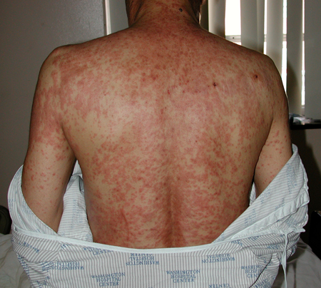
Bullous impetigo
This condition is highly contagious and is a major concern for schools and playgroups. Friction blisters and eczema may be complicated by secondary impetigo, which could evolve into cellulitis and sepsis. Topical antibiotics (e.g., mupirocin or fusidic acid) are effective, but resistance to these agents is not uncommon. Systemic antibiotics may be necessary.
Staphylococcal scalded skin syndrome
Exfoliative toxin in patients with bullous impetigo may disseminate and cause generalised staphylococcal scalded skin syndrome (SSSS) in immunodeficient states. Rupture of bullae can lead to rapid desquamation with impairment of thermo-regulation, and fluid and electrolyte balance. Mortality in children with SSSS is 4% despite fluids, correction of electrolyte imbalances, and antibiotics.[34] Adult patients with impaired renal function can accumulate the toxin and develop clinical symptoms.[34]
Pemphigus vulgaris
Widespread cutaneous involvement may result in death from sepsis or fluid and electrolyte imbalance.[1] Prompt diagnosis and treatment, initially with high doses of prednisolone and antibiotics to control any signs of infection, are often needed in these patients.
Paraneoplastic pemphigus
This condition must be considered in patients who present with intractable stomatitis in the context of an underlying malignancy or immunoproliferative process. Patients may develop progressive respiratory failure due to autoimmune lung injury from bronchiolitis obliterans and secondary pneumonia. The disease course is progressive and often fatal within 2 years.[26]
Pemphigoid
Characterised by large, tense, subepidermal bullae in the groin, axillae, trunk, thighs, and flexor surfaces of the forearms. Some patients present with localised disease, typically on the shins.[1] In addition to bullae, there are often erythematous or urticarial plaques. Children with pemphigoid more commonly have involvement of the face, hands, feet, and genitalia.[35] Certain medications have been implicated in bullous pemphigoid, including furosemide, captopril, non-steroidal anti-inflammatory drugs, penicillamine, systemic antibiotics, and immune checkpoint inhibitors.[21][27][31] Dipeptidyl peptidase 4 inhibitors, particularly vildagliptin, may be associated with the development of bullous pemphigoid in patients with diabetes.[36][37][38] Corticosteroids, either topical or systemic, are first-line treatment.[Figure caption and citation for the preceding image starts]: Bullous pemphigoid (BP)Courtesy of Daniel Eisen, MD [Citation ends].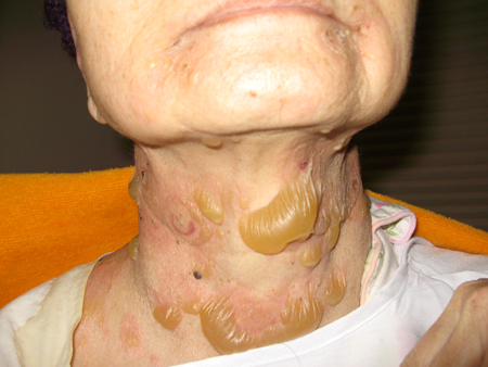 [Figure caption and citation for the preceding image starts]: Bullous pemphigoid (BP)Courtesy of Daniel Eisen, MD [Citation ends].
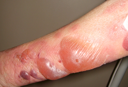
Pemphigoid gestationis
Pemphigoid gestationis carries a risk of low birth weight and in rare cases fetal demise. Pemphigoid gestationis can recur with subsequent pregnancies.[1] Treatment hinges on the use of topical or systemic corticosteroids.
Mucous membrane pemphigoid
May involve oral, ocular, nasal, pharyngeal, laryngeal, oesophageal, and/or anogenital mucous membranes.[29] To prevent irreversible conditions such as blindness and stenosis of laryngeal and pharyngeal mucosa, early diagnosis and treatment is essential. Topical and systemic corticosteroids are used, as well as other immunosuppressive and anti-inflammatory agents.
Mastocytosis
Risk of anaphylaxis is elevated in mastocytosis due to the increased release of anaphylactic mediators from an excess number of mast cells.[39] It is imperative that patients with mastocytosis are educated about avoiding physical stimuli (e.g., pressure, friction, extremes of temperature) and chemical degranulators of mast cells. Treatment is with antihistamines and avoidance of mast cell triggers.
Stevens-Johnson syndrome
Depending on the body surface area involved, there is an increased risk of death from sepsis or fluid and electrolyte imbalance with Stevens-Johnson syndrome. Ocular mucosal involvement mandates an urgent ophthalmological consultation to minimise permanent ocular damage, including blindness. This can develop over weeks and months, and requires treatment and monitoring. Treatment involves supportive care if there is respiratory, renal, or other organ involvement. Patients are often hospitalised and may require systemic corticosteroids or intravenous immunoglobulin. In drug-induced cases, the first-line therapy is to stop the offending medication.
Toxic epidermal necrolysis
In toxic epidermal necrolysis, life-threatening mucocutaneous reactions may be induced by medications, infections, or malignancy, but could also have no clear trigger. The disease presents acutely with generalised erythema followed by desquamation involving >30% of the skin surface.[1][21] Ocular mucosal involvement mandates an urgent ophthalmological consultation to minimise permanent damage, including blindness; this can develop over weeks and months, and requires regular treatment and monitoring. Optimal wound care, nutrition, critical care, and pain management are provided in an intensive care unit or specialised burns unit. Treatments should be initiated on a case-by-case basis, depending on individual patient presentation.[40][41][Figure caption and citation for the preceding image starts]: Toxic epidermolytic necrolysis (TEN)Courtesy of Daniel Eisen, MD [Citation ends]. [Figure caption and citation for the preceding image starts]: Toxic epidermolytic necrolysis (TEN)Courtesy of Daniel Eisen, MD [Citation ends].
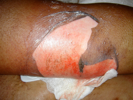
Epidermolysis bullosa acquisita
In epidermolysis bullosa acquisita, the mucosal membranes of the conjunctiva, oral cavity, larynx, and oesophagus may be affected, in addition to the skin. The disease may mimic mucous membrane pemphigoid. Progressive and recurrent disease can result in permanent complications similar to those seen in mucous membrane (cicatricial) pemphigoid, such as blindness and oesophageal strictures.[27] Mild disease may be treated with dapsone or colchicine; more severe disease often requires cyclophosphamide, rituximab, intravenous immunoglobulin, or ciclosporin.
Use of this content is subject to our disclaimer