Images and videos
Images


Legg-Calvé-Perthes' disease
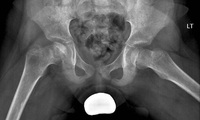
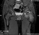
X-ray of stage 3 showing re-ossification
From the personal collection of Dominique Knight
See this image in context in the following section/s:
Legg-Calvé-Perthes' disease
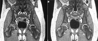
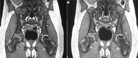
MRI scans of the same patient taken 8 years apart showing Perthes' disease of the left femoral epiphysis and proportional increase in cartilage thickness compared to the right side. Later scan (image on right) starting to show uptake
From the personal collection of Dominique Knight
See this image in context in the following section/s:

Legg-Calvé-Perthes' disease
Original Herring classification: Group A, no involvement of the lateral pillar which retains the original height with no density changes; Group B, lateral pillar shows lucency and a loss of height, not exceeding 50% of the original; Group C, characterises the lateral pillar with increased lucency and collapse of over 50% of the original height
© 1996 American Academy of Orthopaedic Surgeons. Reprinted from: J Am Acad Orthop Surg 1996;4:9-16, with permission
See this image in context in the following section/s:
Legg-Calvé-Perthes' disease
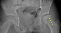
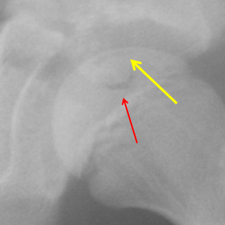
X-ray of stage 2 showing a subchondral fracture line (yellow arrow) and fragmentation (red arrow)
From the personal collection of Dominique Knight
See this image in context in the following section/s:
Legg-Calvé-Perthes' disease
Stulberg classification and prognosis of future hip arthritis
From the personal collection of Jwalant S. Mehta, MS (Orth), MCh (Orth), FRCS, FRCS (Orth)
See this image in context in the following section/s:
Legg-Calvé-Perthes' disease
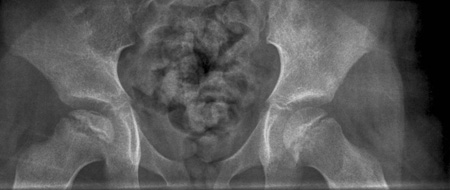
AP radiograph of a patient with Perthes' disease
From the personal collection of Jwalant S. Mehta, MS (Orth), MCh (Orth), FRCS, FRCS (Orth)
See this image in context in the following section/s:
Legg-Calvé-Perthes' disease
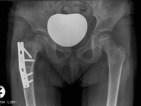
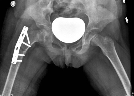
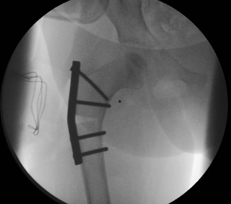
Postoperative radiograph at 4 weeks
From the personal collection of Jwalant S. Mehta, MS (Orth), MCh (Orth), FRCS, FRCS (Orth)
See this image in context in the following section/s:
Legg-Calvé-Perthes' disease
Catterall classification: Group I shows anterior head involvement (hatched area), no sequestrum, no collapse of the epiphysis. Group II shows the anterior head involvement with a clearly demarcated sequestrum. Group III shows that only a small part of the epiphysis is not involved. Group IV shows a total head involvement
© 1996 American Academy of Orthopaedic Surgeons. Reprinted from: J Am Acad Orthop Surg. 1996;4:9-16, with permission
See this image in context in the following section/s:

Legg-Calvé-Perthes' disease
Frog lateral radiograph of a patient with Perthes' disease
From the personal collection of Jwalant S. Mehta, MS (Orth), MCh (Orth), FRCS, FRCS (Orth)
See this image in context in the following section/s:

Legg-Calvé-Perthes' disease
Postoperative radiograph at 3 months
From the personal collection of Jwalant S. Mehta, MS (Orth), MCh (Orth), FRCS, FRCS (Orth)
See this image in context in the following section/s:

Legg-Calvé-Perthes' disease
MRI showing partial collapse of the left femoral head with areas of necrosis
From BMJ Case Reports http://casereports.bmj.com/cgi/content/full/2009/jan08_1/bcr2007132811Copyright © 2011 by BMJ Publishing Group Ltd
See this image in context in the following section/s:
Legg-Calvé-Perthes' disease
Surgical containment
From the personal collection of Jwalant S. Mehta, MS (Orth), MCh (Orth), FRCS, FRCS (Orth)
See this image in context in the following section/s:
Use of this content is subject to our disclaimer