Aetiology
Torsional variations of the lower extremity are rotational malalignments that fall within 2 standard deviations (SDs) of the mean.[2]
Torsional deformities are defined as rotational abnormalities outside the normal range of 2 SDs. Deformities are caused by:
Genetics: similar to other congenital deformities.
Intrauterine positioning: abnormal shaping of structures caused by mechanical forces. These are deformations distinguished from dysplasias (which are genetic or cellular abnormalities that result in structural abnormalities).
Muscle action: abnormalities in muscle function and tone (e.g., cerebral palsy, myelomeningocele, polio). These strongly affect torsional development during growth.[13][16][17][18][19][20]
These factors can cause torsional deformities at any of 3 levels of the lower extremity: foot, tibia, and femur.[2] They often exist together. Excessive pelvic rotation can also contribute to gait abnormalities, especially in children with cerebral palsy. Most rotational problems in normal infants and children are physiological or postural problems. Medial femoral torsion (i.e., excessive femoral anteversion) is an abnormal amount of version between the axes of the femoral neck and the femoral condyles. Increased femoral anteversion allows increased hip medial rotation.[21] Medial femoral torsion (MFT) is associated with sitting in the W position, ligamentous laxity, and conditions such as developmental dysplasia of the hip, cerebral palsy, and hypotonia.[22][Figure caption and citation for the preceding image starts]: Photo of a child sitting in the W positionFrom the collection of Tamir Bloom, MD [Citation ends].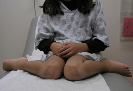 MFT is the most common cause of in-toeing in children older than 6 years.[23] Medial rotation of the hip decreases through adolescence, with correction of in-toeing by late childhood in most.[1][24]
In-toeing caused by medial tibial torsion (MTT) most often is a variation during normal development. MTT is the most common cause of in-toeing in children younger than 5 years.[23] However, many children, regardless of age, will demonstrate a combination of MTT and MFT as the cause of their in-toeing.[23] In-toeing caused by metatarsus adductus is hypothesised to be due to abnormal intrauterine mechanical forces acting on the foot. In-toeing seen in club foot is a congenital deformity with a foot that is turned inward and points downward. It may be idiopathic or associated with myelomeningocele, arthrogryposis, and other syndromes. Although a genetic predisposition exists for torsional abnormalities, no single cause has been identified. Skew foot may be caused by improper casting of metatarsus adductus. Most cases occur due to an unknown cause.
Out-toeing caused by lateral femoral torsion is commonly from persistent hip abduction contractures that develop in utero. As with medial tibial torsion and metatarsus adductus, abduction contractures usually improve during the first year of walking. One in 5 fetuses is positioned with the lower limbs in external rotation, leading to lateral tibial torsion and calcaneovalgus feet. Slipped capital femoral epiphysis is a displacement of the proximal femoral epiphysis around the axis of the femoral metaphysis. This results in an external rotation deformity of the affected lower extremity.
Brain-injured children (e.g., cerebral palsy) have loss of selective muscle control, difficulties with balance, and abnormal muscle tone (primary abnormalities). Torsional deformities develop because the effects of brain injury impose abnormal forces on both muscle and bone. Skeletal deformities may emerge gradually, directly proportional to the rate of skeletal growth.
Pathophysiology
In-toeing
Medial (internal) tibial torsion (MTT): if severe, gait may be compromised by disrupting shock absorption of the foot during loading response and may compromise limb clearance in the swing phase.[25][26] Quantitative gait analysis studies of patients with extreme in-toeing gait because of MTT have demonstrated increased varus loading of the knee during stance phase, in addition to other primary and compensatory gait deviations.[26][27][28] Longstanding severe MTT, related dynamic gait deviations, and associated abnormal loading may be associated with degenerative arthritis of the knee in adults, although the nature of this association is not clear.[26][27][29][30][31][32]
Medial (internal) femoral torsion (MFT): in utero femoral anteversion is about 55°. As a child begins to walk, the hip extends and the anterior iliofemoral ligament stretches over the proximal femur, pushing it backwards.[33] This mechanism remodels the proximal femur. Femoral anteversion measures, on average, 10° to 15° by adulthood. Ligamentous laxity or neuromuscular conditions may have insufficient tension to bring about normal remodelling, resulting in persistent fetal alignment.[34] Limited remodelling of the proximal femur is common in cerebral palsy due to delayed onset of walking and abnormal muscle forces. When walking does begin, the hips are flexed. These factors weaken the iliofemoral ligament tension, promoting anteversion. To seat the femoral head in the acetabulum, children walk with the femur in internal rotation, which results in excessive internal rotation of the entire lower limb.[33] Increased femoral anteversion can occur from torsional loading of the femur in the W sitting position. This position may place an increased torsional load on the femur.
Metatarsus adductus: abnormalities in bony anatomy, such as a deformity of the medial cuneiform, and muscle imbalance or contracture have been suggested.
Skew foot: this is a complex deformity of the foot composed of forefoot adduction, midfoot abduction, and hindfoot valgus.
Club foot: this is a rigid deformity consisting of ankle equinus, heel varus, and midfoot and forefoot adductus.
Blount's disease: infantile type (0-4 years old) is a non-physiological form of genu varum and medial tibial torsion caused by a growth disorder of the medial proximal tibia. Deformity is restricted to the proximal tibia and is associated with early walking and obesity. Adolescent type (after age 9 years) shares similar pathophysiology with the infantile type with excessive loads on a varus knee resulting in abnormal growth of the proximal tibial physis. Unlike the infantile type, deformity may also involve the distal femur.
Out-toeing
Lateral (external) tibial torsion (LTT): if severe, may compromise the stability and lever function of the foot during mid and terminal stance phase of the gait cycle.[35]
Lateral (external) femoral torsion: intrauterine positioning of the legs causes lateral rotation hip (abductor) contractures, which result in increased hip external rotation relative to internal rotation and apparent out-toeing in infancy.[3][36] This usually resolves with early walking.[37]
Slipped capital femoral epiphysis: the femoral neck slips anteriorly and laterally through the physis (growth plate), resulting in an apparent posterior shift of the epiphysis. Increased hip external rotation, decreased hip internal rotation, and increased foot progression angle, as well as other anatomical, clinical, and gait abnormalities, are associated with deformity of the proximal femur. Functional abnormalities may improve after treatment and recovery and are influenced by severity of residual anatomical deformity.[38][39]
Flexible flat feet: this is a normal variation of foot alignment in early childhood, with spontaneous resolution in the first decade of life in nearly all children. Arch height is determined by the bone-ligament complex of the foot and is not influenced by shoe wear.
Effect of torsion on the patellofemoral joint
Bone alignment is one factor that may contribute to patellofemoral joint mechanics.[40] Abnormal limb alignment in the axial plane may alter the balance of body-weight transfer to the ground, overloading biological tissues (ligaments, articular cartilage, bone, muscle, and tendon).
Greater-than-normal internal knee rotation during gait, due to either excess femoral anteversion or lateral tibial torsion, or both, increases the lateral patellofemoral joint compression forces and the strain on the medial patellofemoral ligaments. If severe enough, the physiological threshold of biological tissues may be exceeded, producing anterior knee pain, patellar instability, or knee osteoarthrosis in later life.[29][30][31][41][42][43] This is called miserable malalignment syndrome.[44]
Deformities in children with neuromuscular disease
Torsional deformities arise because of abnormal forces acting on the growing skeleton by the effects of the primary brain injury.[33] Bones fail to mould, remodel, or model normally with growth.
Torsional malalignment is a type of lever-arm dysfunction. Improper transverse plane orientation of the bone causes pathological gait. Bones are levers and rigid bodies on which a load and force act. Their purpose is to produce a mechanical advantage over the load or rapid motion of the load. Malrotated levers produce pathological dysfunction by:
Reduction of the magnitude of moments, reducing the ability of muscles to rotate joints of the limb. A moment is the rotatory impetus occurring at a pivot point (fulcrum) generated by forces acting at a distance of a lever arm.
Introduction of secondary moments that alter leverage necessary for normal gait and promoting further deformities of the long bones by applying abnormal stresses on plastic, growing bones.
Classification
Direction of deformity[1]
Clinical assessment of the direction of lower-extremity rotational alignment is classified as follows.
In-toeing: the long axis of the foot is internally (medially or towards midline) rotated to the line of progression when walking or running.
Out-toeing: the long axis of the foot is externally (laterally or away from midline) rotated to the line of progression when walking or running.
Use of this content is subject to our disclaimer