Images and videos
Images
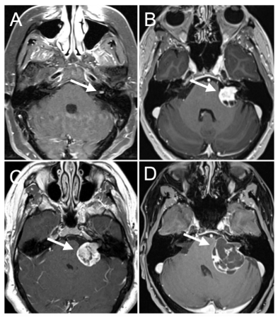
Vestibular schwannoma
Contrast-enhanced T1-weighted axial MRI studies demonstrating various sizes of left-sided sporadic vestibular schwannoma. (A) small intracanalicular vestibular schwannoma; (B-C) medium-sized vestibular schwannomas; (D) large cystic vestibular schwannoma
From the personal collection of Michael J. Link and Matthew L. Carlson, Mayo Clinic, Rochester, MN; used with permission
See this image in context in the following section/s:
Use of this content is subject to our disclaimer