Investigations
1st investigations to order
MRI head
Test
MRI with gadolinium is the most sensitive and specific imaging modality. Used in the diagnosis of gliomas, before and after resection surgery, and as a surveillance method to detect the progression of previously diagnosed and treated tumours.[21][22][24][26][27][28][29]
[Figure caption and citation for the preceding image starts]: T2-weighted MRI without (A) and with (B) contrast demonstrating a pontine gliomaFrom the collection of Karine Michaud, University of California, San Francisco [Citation ends].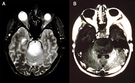 [Figure caption and citation for the preceding image starts]: MRI: T2 and T1 post-contrast, demonstrating a tectal glioma (grade 2)From the collection of Karine Michaud, University of California, San Francisco [Citation ends].
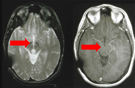 [Figure caption and citation for the preceding image starts]: MRI demonstrating a right temporal glioblastoma (grade 4)From the collection of Karine Michaud, University of California, San Francisco [Citation ends].
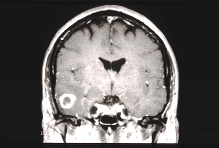 [Figure caption and citation for the preceding image starts]: MRI demonstrating a right frontal grade 2 diffuse astrocytomaFrom the collection of Karine Michaud, University of California, San Francisco [Citation ends].
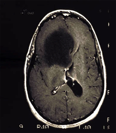 [Figure caption and citation for the preceding image starts]: MRI demonstrating a cerebellar pilocytic astrocytoma (grade 1)From the collection of Karine Michaud, University of California, San Francisco [Citation ends].
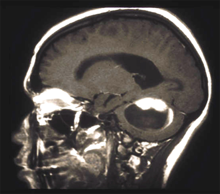
Result
area of hypointensity on T1 sequences and hyperintensity on T2 sequences; contrast enhancement with gadolinium injection, most frequently in high-grade gliomas
ophthalmological evaluation; visual field testing
Test
Ordered if there are specific reports from the patient and/or specific findings on physical examination or evidence of compression of optic chiasm on MRI.
Result
possible visual field defect
CT head
Test
Ordered if the presentation is acute and there is a need to rule out other processes such as herniation, stroke, and haemorrhage.
Also used when MRI is unavailable or contraindicated.
Useful imaging modality for demonstrating calcifications (often seen in oligodendroglioma) and haemorrhage.
Result
area of hypodensity; enhancement with contrast depending on type and grade of the tumour; hyperdensity if calcification or haemorrhage present
brain MR spectroscopy
Test
Used to help differentiate malignant from non-malignant lesions. MR spectroscopy can also be used to differentiate between true tumour progression and treatment-related imaging changes or pseudo-progression in patients with suspected progressive glioblastoma.[26][27]
Result
lactate peak correlates with areas of anaerobic metabolism; N-acetylaspartate (NAA) peak reduced and choline peak elevated in malignant lesions; ratio chol/NAA ≥2
brain perfusion MRI
Test
Provides quantitative assessment of regional cerebral blood flow and cerebral blood volume, reflecting the magnitude of angiogenesis in a tumour, thus helping to differentiate tumour progression from radiation-induced changes in enhancing areas of previously treated tumours.[21]
Result
perfusion elevated in malignant lesions
histopathology
Test
Provides diagnostic information that is integrated with molecular analyses to determine tumour type and grade.
Uses tissue obtained from surgical resection or from stereotactic or open biopsy. One of the limitations of the stereotactic biopsy is the sampling error in diffuse astrocytoma (different areas of the tumour can show different histological grades).
Result
presence of abnormal cells
molecular analyses
Test
Essential for accurate classification and to guide subsequent treatment decisions. Molecular markers that should be investigated (depending on clinical context) include isocitrate dehydrogenase (IDH) 1 and IDH2 mutation, 1p/19q-codeletion, ATRX mutation, TP53 mutation, MGMT promoter methylation status, EGFR amplification, chromosome 7 gain, chromosome 10 loss, TERT promoter mutation, and histone H3 mutation.[1][21][22][24][30]
Result
one or more markers detected
Investigations to consider
pituitary hormones tests
Test
Ordered if there is a known hypothalamic or pituitary region mass, if the patient has received cranial radiation involving the pituitary gland, or if the patient has symptoms or signs of hypopituitarism.
Result
may demonstrate hypopituitarism
Emerging tests
2-hydroxyglutarate-targeted magnetic resonance spectroscopy
Test
2-hydroxyglutarate, an oncometabolite, is produced by isocitrate dehydrogenase (IDH) mutant enzymes.
Optimised magnetic resonance spectroscopy can determine IDH-mutational status by detecting 2-hydroxyglutarate directly.[36]
Result
IDH mutation is associated with the accumulation of 2-hydroxyglutarate in the tumour.
Use of this content is subject to our disclaimer