Images and videos
Images


Atrioventricular block
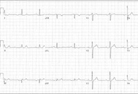
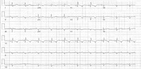
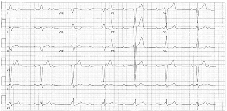
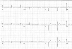
Baseline ECG for a patient with third-degree AV block
Courtesy of Dr Susan F. Kim, Dr John F. Beshai, and Dr Stephen L. Archer; used with permission
See this image in context in the following section/s:
Atrioventricular block
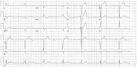
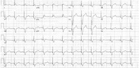
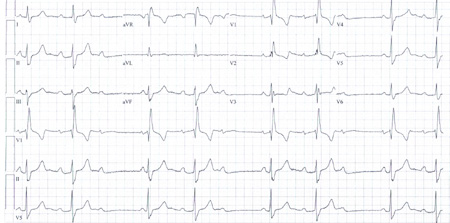
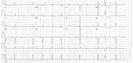
Third-degree heart block: right bundle-branch block escape
Courtesy of Dr Sanjiv Petkar; used with permission
See this image in context in the following section/s:
Atrioventricular block
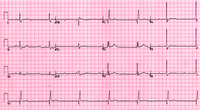
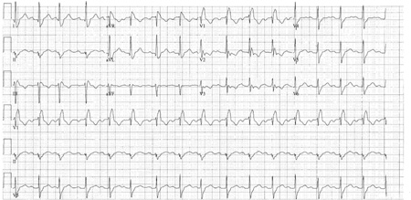
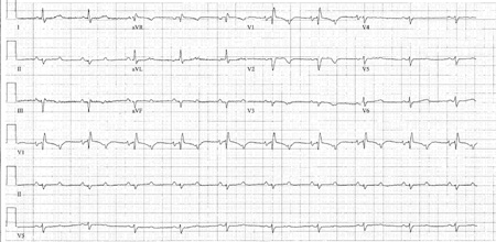
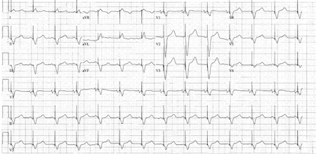
Third-degree heart block: right bundle-branch block escape
Courtesy of Dr Sanjiv Petkar; used with permission
See this image in context in the following section/s:
Atrioventricular block
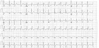
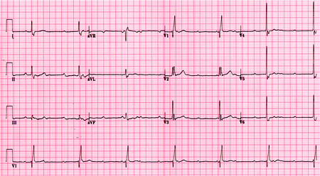
Third-degree AV block
Courtesy of Dr Susan F. Kim, Dr John F. Beshai, and Dr Stephen L. Archer; used with permission
See this image in context in the following section/s:
Atrioventricular block
Type I second-degree AV block. This figure demonstrates typical features of the AV Wenckebach block, including progressively shortening R-R intervals as the P-R intervals lengthen; the figure also shows grouped beating, which is also typical for AV Wenckebach block
Courtesy of Dr Susan F. Kim, Dr John F. Beshai, and Dr Stephen L. Archer; used with permission
See this image in context in the following section/s:
Atrioventricular block
2:1 AV block
Courtesy of Dr Susan F. Kim, Dr John F. Beshai, and Dr Stephen L. Archer; used with permission
See this image in context in the following section/s:
Atrioventricular block
Third-degree AV block
Courtesy of Dr Susan F. Kim, Dr John F. Beshai, and Dr Stephen L. Archer; used with permission
See this image in context in the following section/s:
Atrioventricular block
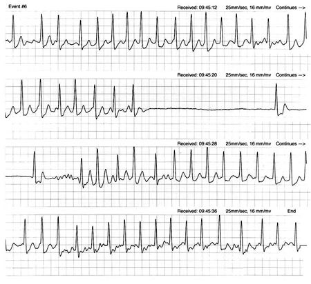
Tachy-brady syndrome due to sinus node disease. Ventricular rate is slow intermittently, but AV block is not seen
Courtesy of Dr Sanjiv Petkar; used with permission
See this image in context in the following section/s:
Atrioventricular block
First-degree AV block
Courtesy of Dr Susan F. Kim, Dr John F. Beshai, and Dr Stephen L. Archer; used with permission
See this image in context in the following section/s:
Atrioventricular block
Type II second-degree AV block
Courtesy of Dr Susan F. Kim, Dr John F. Beshai, and Dr Stephen L. Archer; used with permission
See this image in context in the following section/s:
Atrioventricular block
Patient with 2:1 AV block, status post permanent pacemaker placement
Courtesy of Dr Susan F. Kim, Dr John F. Beshai, and Dr Stephen L. Archer; used with permission
See this image in context in the following section/s:
Use of this content is subject to our disclaimer