Approach
The diagnosis of volume depletion is clinical, based on the history and physical examination. Symptoms can range from thirst (with mild depletion) to irreversible shock and death in severe cases. Experienced clinicians will be able to do a volume status- focused physical examination despite the challenges of an uncooperative, agitated, or crying child. Presenting signs and symptoms of tachycardia and irritability can be mistakenly attributed to anxiety or behavioural issues. If a child is agitated or anxious for the initial physical examination, a provider can complete serial examinations or monitor continuous vital signs remotely to better assess the clinical status of the patient.
There is no specific laboratory test for volume depletion, and diagnosis is based on clinical findings and history, particularly focused on oral intake and volume of stool and urine output. Patients who present with inconsistent history, severe volume depletion, or who do not respond to initial fluid resuscitation are a small subset of patients in whom diagnostic testing should be considered. Use of a clinical scale or scoring system may improve diagnostic accuracy.[17] Some laboratory tests may prove useful in directing therapy for moderate to severe illness.[18][19]
Presence of risk factors
Strongly associated risk factors for volume depletion in children include:
Vomiting and/or diarrhoea (most commonly associated with gastroenteritis).
A history of trauma: haemorrhage is generally quite obvious, but the higher incidence of blunt trauma, the large size of a young child's internal organs and head relative to skeletal size, and the child's inability to communicate localised pain are all challenges to recognising haemorrhagic losses in children. Additionally, non-accidental trauma (physical abuse) is too often not considered early in the assessment of a child with altered mental status or non-specific complaints. Non-accidental trauma may be associated with significant intracerebral haemorrhage, which in a very young infant can lead to haemorrhagic shock and hypovolaemia.
A history of burns to over 10% of the body surface area causes significant losses through the disrupted skin barrier.
History or symptoms of diabetes mellitus: children without a previous diagnosis or with delayed presentation to care, very young children, and adolescents are most likely to present with significant volume depletion from hyperglycaemia and resultant glycosuria.[20]
A history of poor oral intake may be present. A child who is refusing to drink due to nausea, pain, altered mental status, or other reasons is at risk for becoming dehydrated, and consequently volume-depleted.
A history of diuretic use. Diuretics promote additional excretion of free water from the kidney, thus limiting the kidney’s ability to compensate for volume depletion by retaining more water and solute, which can lead to volume depletion.
A history of vigorous and prolonged exercise may also be present: adolescent athletes exercising heavily in high ambient humidity and temperature can have significant losses, which, if not replaced by frequent hydration with appropriate fluids, leads to volume depletion.[16][21]
History of recent symptoms
Thirst: a history of thirst prompts investigation of a hyperosmolar state, as seen with relative dehydration and hypernatraemia.
Vomiting occurs frequently in both gastroenteritis and diabetic ketoacidosis (DKA; secondary to the nausea caused by ketosis) and may prevent oral rehydration therapy. It is important to ask when the last intake occurred, and what was given, so therapy can be planned.
Diarrhoea (>3 watery stools/day) characterises gastroenteritis. The onset, quantity, frequency, and presence of blood or mucus should be investigated. Blood and mucus in the stool suggest bacterial pathogens.
Abdominal pain is common in gastroenteritis, intra-abdominal haemorrhage, and small-bowel obstruction.
Urinary output is high in cases of excess renal losses (diabetes insipidus, DKA, tubular defects), but is appropriately low in other conditions of hypovolaemia.
Fever may be seen in uncomplicated gastroenteritis but also suggests more severe, invasive disease. It is associated with increased insensible losses, associated respiratory losses through tachypnoea, and increased metabolic rate. It is a risk factor for sepsis.[22]
Vital signs and initial glucose check
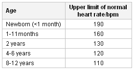
Tachycardia is a reliable indicator and may occur even with mild volume depletion. Children with hypernatraemic dehydration may have relative preservation of intravascular volume compared with those with hyponatraemic dehydration, and therefore have a somewhat slower heart rate despite an equivalent overall volume loss. Infants and young children with hypovolaemia maintain adequate cardiac output primarily through an increase in heart rate, due to a developmentally limited capacity to augment stroke volume. [Figure caption and citation for the preceding image starts]: Upper limits of normal heart rate by ageTable created by the BMJ Group. Based on data from Ngo NT, et al. Clin Infect Dis. 2001;32:204-213 [Citation ends].

BP: unless volume depletion has progressed to severe shock, BP is maintained or even slightly elevated. Children can preserve a normal BP over a much wider range of volume depletion than adults. Large volume losses are generally necessary to manifest as changes in BP. In children, low BP is a late and ominous sign. Tables of normal values for BP in children have been published. Clinical practice guideline for screening and management of high blood pressure in children and adolescents Opens in new window
Respiratory rate and depth: hyperpnoea or tachypnoea is seen as a compensatory response to a metabolic acidosis. This occurs with lactic acidaemia from decreased tissue perfusion and ketonaemia in DKA.
Temperature is commonly elevated with infectious illness, burns, heat stress, and sepsis. A low core temperature can indicate significant haemorrhage, sepsis (particularly in a young infant), and shock. A peripheral skin temperature that is notably lower than the patient's central temperature is a result of increased systemic vascular resistance and indicates a state of compensated shock in hypovolaemia.
Glucose test strip: hypoglycaemia occurs frequently in ill infants due to higher metabolic rates and lower glycogen stores. Rapid bedside blood glucose measurements should be obtained in all young children presenting with altered mental status and signs of volume depletion, and should be confirmed with serum glucose to rule out false elevation that may mask hypoglycaemia. Hypoglycaemia should be rapidly addressed. Patients presenting with volume depletion from new-onset diabetes are hyperglycaemic.
General examination
Mental status, activity level assessment: this provides critical diagnostic information. Infants and small children who are inconsolable or listless, or do not seem to resist invasive or uncomfortable procedures, should be assumed to have serious/deteriorating illness.
Mucous membranes: dry or tacky mucous membranes are seen with hypovolaemia; pallid mucous membranes suggest blood loss with anaemia.
Capillary refill: classically, volume depletion is associated with a prolonged capillary refill time (>3 seconds). This is most likely to be true in the setting of gradual volume depletion, as is seen in gastroenteritis. A meta-analysis concluded that the 3 most useful clinical findings in a child with volume depletion and dehydration were prolonged capillary refill time, decreased skin turgor, and abnormal respiratory pattern.[23] However, in burns, anaphylaxis, and sepsis, capillary refill time may not be prolonged (<3 seconds). Therefore, determining capillary refill time is not a reliable clinical test in all cases.
Skin turgor can be notably affected in severe cases of volume depletion, particularly those associated with hypernatraemia or hyperosmolarity. A doughy consistency is reported in hyponatraemic states. Because children have more skin elasticity than adults, this is often a relatively late sign in the progression of volume depletion. Assessing skin turgor by pinching a small fold of skin on the abdomen adjacent to the umbilicus and observing recoil is recommended. In the setting of severe acute malnutrition, however, skin turgor is not a reliable sign of volume depletion. In these patients, skin tenting occurs due to the loss of subcutaneous fat rather than solely volume depletion.
Bruises or signs of neglect: children presenting with hypovolaemia from internal bleeding as a result of non-accidental trauma may have evidence of prior trauma or general neglect. Importantly, these signs may be completely absent. The lack of external findings is not sufficiently reassuring to preclude further investigation.
Abdominal examination: abdominal tenderness is common in gastroenteritis, intra-abdominal haemorrhage, and small-bowel obstruction. Active bowel sounds are heard in gastroenteritis. Bowel sounds are diminished in sepsis, hypokalaemia, some kinds of abdominal trauma, and generalised severe illness.
Commonly used indicators to determine level of volume depletion
Mild to moderate volume depletion
Preserved normal mental status
Haemodynamic stability
Mildly altered vital signs (e.g., minor degree of tachycardia)
Severe volume depletion
Altered mental status
Sustained tachycardia
Haemodynamic instability
WHO: Manual for the management of diarrhoea for physicians and senior health workers
Classifies symptoms of volume depletion in children presenting specifically with diarrhoeal illnesses.[14] Symptoms and signs allow for placement of children into 1 of 2 categories according to severity of volume depletion.
Mild to moderate dehydration (5% to 10% loss of total body water)
General: restless, irritable
Eyes: sunken
Thirst: thirsty, drinks eagerly
Skin pinch: goes back slowly
Severe dehydration (>10% loss of total body water)
General: lethargic or unconscious.
Eyes: sunken
Thirst: drinks poorly or cannot drink
Skin pinch: goes back very slowly
American College of Surgeons: Classification of estimated blood loss in children
Classifies estimated blood loss by clinical signs in children.[24] Simplifies presentation to 4 systems in 3 categories (mild, moderate, and severe).
Mild (15% to 30% total blood volume loss)
Cardiovascular system (CVS): tachycardic, weak pulses
Central nervous system (CNS): irritable, confused, agitated
Skin: cool, prolonged capillary refill
Urine output: minimally decreased
Moderate (30% to 45% total blood volume loss)
CVS: tachycardic, thready peripheral pulses, narrow pulse pressure, mild hypotension
CNS: lethargic, minimally responsive
Skin: cyanotic or pallid, extremely prolonged capillary refill
Urine output: minimal
Severe (>45% total blood volume loss)
CVS: tachycardic, absent peripheral pulses, hypotensive, late bradycardia
CNS: comatose
Skin: pale, cold
Urine output: none
Investigations
Urine osmolality and specific gravity: concentration of urine leading to a high urine osmolality is an appropriate physiological response to volume depletion, but may not reliably predict dehydration.[17][25] Infants younger than 6 months are less able to concentrate urine due to renal immaturity and may not have a high specific gravity. Patients with primary renal losses have inappropriately dilute urine in the face of significant volume depletion. Urine is analysed with microscopy and culture for glycosuria, proteinuria, and signs of urosepsis.
Serum electrolytes and blood glucose: differentiating hypernatraemic, isonatraemic, and hyponatraemic volume losses helps to guide subsequent therapy. Low serum bicarbonate is identified in many types of hypovolaemia due to direct losses (e.g., with diarrhoea or poor tissue perfusion from decreased intravascular volume). Glucose levels can be low in young patients with poor intake or markedly elevated in patients with volume depletion accompanying DKA.
Urea/Cr: the ratio is greater than 20:1 with renal hypoperfusion.
FBC: anaemia can be seen in non-acute blood loss, whereas haemoconcentration may be noted in the setting of lost plasma volume, especially burns. Acute haemorrhage without administration of intravenous fluids will often have a relatively preserved haemoglobin level. Leukocytosis or neutropenia is common in the setting of infection.
Blood culture may be performed if sepsis is suspected.
Head and abdominal ultrasound or CT scan: blunt trauma, especially to the head and abdominal viscera, can be a cause of major haemorrhage-associated volume depletion. Altered mental status in a child known to have sustained trauma or in a young infant should prompt investigation for occult bleeding. The history is often hidden in settings of abuse.
Use of this content is subject to our disclaimer