Approach
The diagnosis of a cleft lip with or without cleft palate involves careful neonatal examination immediately after birth. Identification of more subtle deformities such as microform cleft lip and sub-mucous cleft palate require a trained and experienced eye.
Presentation
The possible presence of an oro-facial cleft may be revealed by a routine antenatal ultrasound scan at 18 weeks' gestation. In one systematic review, routine, 2-dimensional ultrasound resulted in a low antenatal detection rate of orofacial clefts in utero. However, 3-dimensional ultrasound showed accurate diagnostic accuracy for detection of antenatal cleft lip and cleft lip with or without cleft palate. Three-dimensional ultrasound did not show any increased accuracy in detecting cleft palate alone.[36] In such cases of positive antenatal ultrasound, involvement of maternal-fetal specialists and genetic counselling is recommended.[37]
A small proportion of infants with any type of cleft palate, including Pierre Robin sequence (triad of cleft palate, microgenia, and glossoptosis), may present with symptoms of severe airway obstruction requiring immediate airway management. Less overt palatal clefting, such as sub-mucous cleft palate, may also present with airway obstruction.
Other presentations include symptoms of feeding difficulties, poor weight gain, hyper-nasal speech, or nasal regurgitation.
Neonatal examination
A thorough head and neck examination begins with assessing and palpating the continuity of the upper lip and nostrils. The various presentations of oro-facial clefting (cleft lip, alveolus, or palate) must be considered as the examination is being carried out.
The upper gum-line and hard palate are palpated for a notched or cleft alveolus and/or palate. In a unilateral or bilateral cleft lip and palate, a complete defect of these structures occurs most commonly under the nostril(s).
A tongue depressor and light are used to view the soft palate back to the uvula.[38] Identification of a bifid or divided uvula may indicate a sub-mucous cleft palate.
The lower lips are inspected for nodular lip pits, and the soft tissues of the cheeks are checked for atypical clefts or auricular remnants found in oculo-auricular-vertebral spectrum (OAVS).
Oro-facial clefts can manifest as unilateral or bilateral; complete, incomplete, or microform (e.g., sub-mucous cleft palate); clefting of the palate in isolation; clefting of the lip with or without the palate; or atypical cranio-facial clefts. The majority of cleft lip deformities are associated with a varying degree of nasal deformity.
Bilateral cleft lip with or without cleft palate [Figure caption and citation for the preceding image starts]: Bilateral cleft lip and palate preoperativelyFrom the collection of Travis T. Tollefson, MD, FACS [Citation ends].
Bilateral cleft lip deformity usually presents with a complete bilateral cleft palate. However, incomplete bilateral and even unilateral cleft palate is also seen.
The configuration of the lateral segments of bilateral cleft lip with or without cleft palate is similar to that of the lateral segment of the unilateral deformity.
The central prolabium and pre-maxilla in bilateral cleft lip and palate are markedly different from those of the unilateral deformity, as there is no muscle in the prolabial skin due to its embryological origin.
Varying severity of clefting of the lip, alveolus, and palate are observed. In complete bilateral cleft lip with or without cleft palate, the pre-maxilla protrudes anteriorly and is totally detached from each maxilla. In incomplete bilateral cleft lip, there is usually some skeletal continuity and very little protrusion of the pre-maxilla and prolabium. If the lateral maxillary alveolar segments are constricted together, a 'locked-out' pre-maxilla is noted. The complete bilateral cleft lip nasal columella is short, contributing to bilateral hooding of the ala and a wide, bulbous de-projected nasal tip.
Unilateral cleft lip with or without cleft palate [Figure caption and citation for the preceding image starts]: Unilateral cleft lip and palate preoperativelyFrom the collection of Travis T. Tollefson, MD, FACS [Citation ends].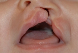
There is extensive variation in the severity of unilateral cleft lip and palate from complete to microform.
In severely wide (>1 cm) complete unilateral cleft lip and palate, there is complete separation of the lip musculature, alveolus, and palate.
An incomplete cleft lip with or without cleft palate extends to more than one quarter of the labial height, and is measured from the normal peak of the upper lip junction between the white and red lip (Cupid's bow) to the bottom of the nostril (nasal sill).
A microform cleft is the most diminutive form of lip cleft.
In an isolated unilateral cleft lip, the base of the nose splays laterally when the infant smiles. The nose and lip on the non-cleft side is characterised by a short vertical white lip height, deficient medial mucosa, deficient columellar skin, caudal septal deflection to the non-cleft side, and a cleft lip nasal deformity with an asymmetrical nasal tip (secondary to lower lateral cartilage dysmorphology).
Isolated cleft palate
The degree of clefting can range from a complete isolated cleft palate to a bifid uvula.
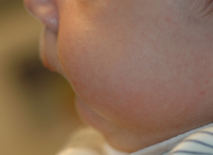
An example of a deformational cleft palate is seen in Pierre Robin sequence, which is characterised by the classic triad of micrognathia, glossoptosis, and an isolated cleft palate. [Figure caption and citation for the preceding image starts]: Lateral view of infant with Pierre Robin sequenceFrom the collection of Travis T. Tollefson, MD, FACS [Citation ends].
Microform cleft lip [Figure caption and citation for the preceding image starts]: Microform cleft lipFrom the collection of Travis T. Tollefson, MD, FACS [Citation ends].
Characterised by dysmorphic features including a minor nasal deformity, philtral groove, indented free mucosal margin, and notched vermilion-cutaneous junction with disruption extending to no more than one quarter of the labial height, measured from the normal peak of the upper lip junction between the white and red lip (Cupid's bow) to the bottom of the nostril (nasal sill).[39]
Isolated sub-mucous cleft palate [Figure caption and citation for the preceding image starts]: Sub-mucous cleft palateFrom the collection of Travis T. Tollefson, MD, FACS [Citation ends].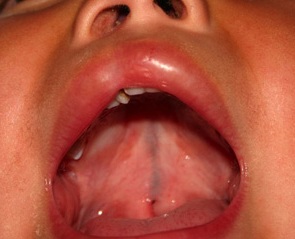
Less overt palatal clefting, such as a sub-mucous cleft palate, may be missed on examination and may instead present with associated symptoms.
Sub-mucous cleft palate is identified by a diastasis of the midline palatal musculature (zona pellucida), a notched hard palate on palpation, and a bifid uvula.
The neonate is also observed feeding at the breast or with a bottle for detection of difficulty in latching on and creating adequate suction. Breastfeeding is possible with an isolated cleft lip. However, neonates with a cleft palate often cannot produce the negative pressures necessary on suction.
Specialist evaluation
If an oro-facial cleft is diagnosed, the infant is evaluated for the presence of associated syndromic features.
Consultations with a geneticist and cleft surgeon allow evaluation of the presence of cardiac defects, limb deformities, microgenia, or renal malformation. Ophthalmological consultation for identification of Stickler syndrome is undertaken in infants with Pierre Robin sequence.
Investigations
Audiology assessment
All newborns with oro-facial clefting receive a screening audiogram. If this is inconclusive, an auditory brain stem response (ABR) test to assess for the presence of associated hearing loss is done by an audiologist. This is useful in the assessment of congenital sensorineural hearing loss. A complete audiological evaluation is indicated in children whose screening audiogram is abnormal.[40]
Imaging
In addition to an audiology assessment, vertebral spine x-rays and renal ultrasound are requested if hemifacial microsomia is present.
Chromosomal analysis
Fluorescence in-situ hybridisation (FISH) is undertaken in order to detect the presence of a genetic disorder (e.g., velocardiofacial syndrome, which results from a micro-deletion of 22q11.2, in infants with isolated cleft palate).
Use of this content is subject to our disclaimer