Investigations
1st investigations to order
chest x-ray
Test
Initial imaging modality of choice in suspected foreign body aspiration.[4]
Radio-opaque foreign bodies are seen in only 2% to 19% of patients because most aspirated objects are radiolucent.
Organic materials, such as meat and vegetables, are difficult to visualise.[35]
The sensitivity of chest x-ray performed in the emergency department for foreign body aspiration has been reported to be 22.6%.[37] False-negative rates vary between 5% and 30% in children, and between 8% and 80% in adults, probably because of differences in the physical properties of the aspirated materials.
Non-specific findings that suggest foreign body aspiration include atelectasis, pneumonia, air trapping, and pneumomediastinum. Bronchiectasis, lung abscess, and empyema are usually late findings.
A normal chest x-ray does not rule out foreign body aspiration, and CT scan or bronchoscopy is warranted for confirmation.
Result
radio-opaque foreign bodies can be visualised
Investigations to consider
CT chest
Test
In children, low-dose multidetector CT (MDCT) scanning with virtual bronchoscopy (VB) can be used to diagnose foreign body aspiration, and to determine the exact location of the obstruction. It has a sensitivity of 92% to 100% and specificity of 80% to 85%.[41][42] False-positives occur because of secretions and endobronchial tumours. In adults, bronchoscopy should be subsequently performed for confirmation or to diagnose alternative causes of airway obstruction.[Figure caption and citation for the preceding image starts]: CT of the chest with intravenous contrast material, showing complete left lower lobe collapse with a radio-opaque object within the left lower main bronchus surrounded by a halo of airBMJ Case Reports 2008 (doi:10.1136/bcr.06.2008.0013). Copyright 2008 BMJ Group Ltd [Citation ends].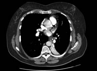
Result
foreign body can be seen in the airway lumen; indirect common findings include atelectasis, hyperlucency, and lobar consolidation
bronchoscopy
Test
Rigid bronchoscopy may be considered as the initial diagnostic and therapeutic test in paediatric patients with asphyxia; radio-opaque foreign body; or in the presence of associated unilateral decreased breath sounds, localised wheezing, hyperinflation, or atelectasis.[14][43][44][45] In all other circumstances, flexible bronchoscopy should be performed first for diagnostic confirmation.[14][45]
In stable children with suspected foreign body aspiration, flexible bronchoscopy has been shown to safely confirm the diagnosis and can be used for therapeutic purposes.[5][44][46][47]
In stable adults, flexible bronchoscopy should be used initially to confirm suspected cases of foreign body aspiration and to attempt removal of the foreign body.[4][51][52][53] It is the initial method of choice in patients with cervicofacial trauma and in those on mechanical ventilation. In these patients, rigid bronchoscopy should be reserved as a therapeutic approach rather than as a diagnostic tool.
Result
determines the nature of the foreign body, its location, the degree of airway obstruction, and associated mucosal abnormalities
Emerging tests
ultrasound lung
Test
Point-of-care ultrasound (POCUS) findings have been reported in a prospective, observational, cross-sectional study of children with foreign body aspiration.[56] However, sample size was small.
Prospective direct comparisons of lung ultrasound with chest x-ray are required.
Result
findings in patients with foreign body aspiration may include B-lines, barcode sign, pleural line abnormalities, and consolidation
Use of this content is subject to our disclaimer