Approach
The initial task in assessment of a patient with suspected food allergy is to separate atopic from non-atopic disease, and to distinguish symptoms and signs of minor adverse immune reactions from more severe concerns of anaphylactic response. It should also aim to identify, if possible, a culprit food. Testing for food allergens should be based on and interpreted in the context of the historical and physical findings.
History and examination
Evaluation should begin with detailing the specific signs and symptoms reported by patient or parent, with particular focus on dermatological, respiratory, gastrointestinal, ophthalmic, and severe cardiac or systemic manifestations. Findings that support the diagnosis of a food allergy include:
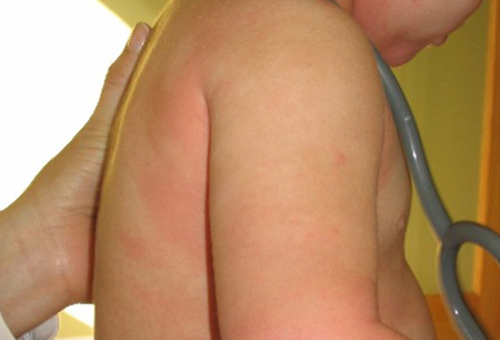
Pruritus, flushing, urticaria, and angio-oedema of the skin[Figure caption and citation for the preceding image starts]: Typical cutaneous findings in food allergy at 30 minutes after ingestion of peanutsFrom the collection of Duke University Medical Center; used with permission [Citation ends].
Sneezing, rhinorrhoea, nasal congestion, metallic taste, hoarseness, stridor, a sense of choking, laryngeal oedema, dyspnoea, tachypnoea, wheezing, coughing, or cyanosis
Nausea, vomiting, abdominal cramping, bloating, and diarrhoea
Conjunctival injection, lacrimation, periorbital oedema
In severe cases, conduction disturbances, tachycardia, bradycardia, arrhythmias, hypotension, and cardiac arrest.
Pertinent clues that support the clinical impression of atopic disease include a family member with food allergy, presence of other allergic disease (e.g., atopic dermatitis, allergic rhinitis, asthma), perinatal transdermal food exposure (e.g., peanut oil), dietary excess or diminished vitamin D, omega-3 polyunsaturated fatty acids or antioxidants, and a paucity of exposure to bacteria and infection. Studies in the UK have shown that if a first-degree family member has peanut allergy, the risk of peanut allergy increases 7 times.[15] Monozygotic twins have been reported to have a 64% concordance rate for food allergy compared with 6.8% among dizygotic twins.[20] Patients with atopic dermatitis, asthma, and allergic rhinitis are more likely to have a food allergy. The presence of asthma is a risk factor for a fatal reaction.[36] Two-thirds of children with atopic dermatitis and food allergy are reactive to egg.[37]
Ninety percent of reactions are caused by milk, egg, peanut, tree nuts, wheat, soya, fish, and shellfish in children, and by peanut, tree nuts, shellfish, fish, and vegetables in adults.[1][38][39] The causative food may often be revealed with careful questioning and consideration of the patient's response.
Has the suspected food allergen been ingested, inhaled, or touched? A specific suspect food should produce symptoms reproducibly nearly every time it is ingested.
How much of the food was ingested at the time of the reaction? IgE-mediated reactions may be triggered by minute amounts, whereas other disorders may require larger amounts.
How soon after exposure to the suspected food allergen did the symptoms occur? IgE-mediated reactions usually occur within 20 minutes of exposure and almost always within 2 hours.
How long did it take for the symptoms to resolve in the past and how was the reaction treated? IgE-mediated symptoms typically resolve within 4 to 12 hours. Reactions may resolve spontaneously or may respond to medical interventions.
Has exercise been associated with the development of symptoms? Food-dependent, exercise-induced anaphylaxis may occur if the food is eaten within 2 to 4 hours before or after exercise.[37]
Were any medications or alcohol ingested in proximity to the reaction? Medications and alcohol are believed to increase the rate of allergen absorption.[40][41][42]
Testing modalities for food allergy
If the initial evaluation is suggestive of food allergy, diagnostic testing should be performed. Testing may begin with either in vitro immunoglobulin (Ig) E immunoassays or skin prick testing. If the assessment is negative (e.g., the patient tolerates the food regularly and has no related symptoms), then diagnostic testing does not need to be performed and food allergy may be ruled out as a cause of the symptoms.[43] If a food has been tolerated in large quantities many times before, it is not likely a relevant allergen, even with a positive test. Commercially prepared extracts of fruits and vegetables are not as predictive because of the lability of the protein. Fresh fruits and vegetables should be used for skin testing.[44]
High sensitivity and low specificity of skin prick and IgE testing for food allergy can yield false positive results, which may lead to elimination diets that are potentially harmful to patients.[43] Effects such as progression to immediate-type allergy, including anaphylactic reactions have been reported.[43][45][46][47] Testing should be performed by an allergy specialist trained in the treatment of rare but potentially life-threatening events. If specific testing falls below values predictive of a reaction by immunoassay testing and by skin testing, or the diagnosis is in question, then a food challenge may be performed. Negative skin tests to foods early in life do not preclude the subsequent development of specific IgE hypersensitivity in later childhood.[22]
Double-blind placebo-controlled food challenges are considered the key test in diagnosing food allergy.[48] These challenges are graded, and there should be an equivalent number of placebo and food steps. If the patient passes this challenge, then an open feeding is performed. If this is tolerated, then food allergy has been excluded.
Investigational studies
Purified or recombinant allergens are used to identify specific IgE sensitisation to proteins within an individual food allergen in component-resolved diagnostics. Some studies have shown an increased ability to predict the likelihood of having a severe allergic reaction to foods like peanut, soy, or hazelnut; however, geographical pollen sensitisation patterns may affect results, and further studies are needed to generalise interpretability.[49][50] The role of component testing continues to evolve.[51]
Atopy patch testing is typically used to identify allergens that cause reactions through delayed contact hypersensitivity where T cells play a major role. Allergenic extract is occluded against intact skin for 48 hours; it is available for investigational use only.[52] Patch testing is well validated for contact dermatitis but not food allergy in general.
Use of this content is subject to our disclaimer