Approach
Diagnosis is usually made via history and physical examination alone; however, supporting laboratory studies may help identify contributing aetiologies, especially when refractory to empirical treatment. Laboratory testing and additional work-up are usually performed only after treatment failure.
History
The first step is a thorough history, with emphasis on identifying medical history, such as:
Chronic skin conditions
Inflammatory bowel disease
Medicines causing xerostomia and immunosuppression
Diabetes mellitus
Denture history
Oral care
Eating disorders
History of total parenteral nutrition
History of acid reflux
Diarrhoea.
It is also important to identify any rash as pruritic or non-pruritic. The patient's age and sex are important to note, as older men are more prone to angular cheilitis.
Oral examination
The second step is a thorough oral examination, checking for any sign of erythema, candidiasis, xerostomia, or abnormal mucosal changes such as palatal erosions. A dental referral is recommended to assess the patient's oral care, teeth, gums, mouth closure, and the lips and tongue.[21][Figure caption and citation for the preceding image starts]: Angular cheilitisFrom the collection of Dr Wanda C. Gonsalves; patient consent obtained [Citation ends].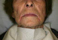 [Figure caption and citation for the preceding image starts]: Angular cheilitisFrom the collection of Dr Wanda C. Gonsalves; patient consent obtained [Citation ends].
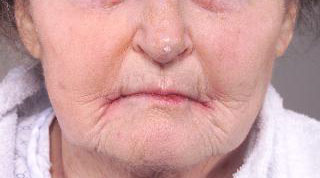
Characteristic painful red fissures may be identified at the corner of the mouth. The 4 possible clinical patterns are:[3]
A single rhagad (painful fissure)
A single deeper and longer rhagad following a skin fold
Several rhagades radiating from the corners of the mouth
Erythema of the skin adjacent to the corners of the mouth without rhagades.
If the examination reveals palatal erythema, then denture-induced stomatitis is usually due to candidiasis.[2][13] If dental caries are apparent, an eating disorder is a possible aetiology. An underlying nutritional deficiency may often be revealed via examination of the tongue.[2][13] The tongue examination may show the following:
Aphthous ulcers or a pale de-papillated tongue in iron deficiency with or without anaemia
A de-papillated glossy red tongue in folate or niacin deficiency
A red atrophic tongue in vitamin B12 deficiency
A reddish-purple de-papillated tongue in riboflavin deficiency.
Riboflavin deficiency also produces smooth, shiny red lips, associated with angular cheilitis; this combination has been called cheilosis.
Angular cheilitis accompanied by alopecia, diarrhoea, and oral ulcerations suggests a zinc deficiency.
An eczematous dermatitis may extend down onto the cheek or chin, as in infective eczematoid reaction, or as a reaction to topical medicines. Severe pruritus would indicate allergic contact dermatitis.
Laboratory evaluation and dental assessment
Laboratory testing and additional work-up are usually performed only after treatment failure. Bacteriological and mycological cultures are recommended, including sampling the mucosal surfaces covered by dentures (i.e., oral mucosa), and the nares, to identify staphylococcal colonisation. Blood samples for FBC, fasting blood glucose, folate, iron, zinc, riboflavin, niacin, and vitamin B12 are indicated to exclude deficiency syndromes.
Angular cheilitis associated with candidiasis resistant to therapy may prompt investigation for an underlying immunological deficiency, such as HIV. Patch testing may be indicated in patients with recalcitrant disease in order to avoid potential allergens, and for recalcitrant, unilateral disease, tissue biopsy is recommended to exclude malignancy.
Use of this content is subject to our disclaimer