History and exam
Key diagnostic factors
common
history of malignancy
In the US, approximately 80% of people with lymphoedema are thought to have disease secondary to cancer or cancer-related treatment, particularly breast cancer.[44]
Lymphoedema occurs in as many as 38% of women following mastectomy for breast cancer with axillary lymph node dissection and radiation.[36]
history of travel to endemic filariasis area
Nematodes such as Wuchereria bancrofti and Brigia malayi cause filariasis by lymph channel obstruction or inflammation. Although broad declines in prevalence have been observed globally, focal areas in Africa and southeast Asia retain higher prevalence rates.[9]
history of previous surgery
history of radiotherapy
Radiation near axillary or groin lymph nodes can cause fibrosis and lymphoedema.[1]
painless unilateral swelling of extremity or genitalia
Secondary lymphoedema is usually unilateral, whereas primary lymphoedema is more frequently bilateral. Defined by swelling that begins distally and progresses proximally.[Figure caption and citation for the preceding image starts]: Primary lymphoedema of the right lower extremityFrom the collection of Dr Arin K. Greene [Citation ends].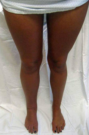 [Figure caption and citation for the preceding image starts]: Secondary lymphoedema: left lower extremity lymphoedema following radiotherapy and lymphadenectomy for Hodgkin's lymphomaFrom the collection of Dr Arin K. Greene [Citation ends].
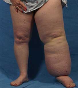
involvement of distal extremity
The hands and feet are almost always involved; hand or foot oedema can occur alone or together with arm or leg involvement.
positive Stemmer's sign
A positive Stemmer's sign (inability to pinch the skin on the dorsum of the second toe between the thumb and index finger) is useful to demonstrate distal involvement.[52] A negative Stemmer's sign does not exclude lymphoedema.
Although this sign was described for the toes, the trained observer can demonstrate this phenomenon on any other part of the body.
Other diagnostic factors
common
history of penetrating trauma to the axilla or groin
Significant penetrating trauma, particularly to the axilla or groin, can cause lymphoedema.
Blunt trauma or minor penetrating trauma does not usually increase the risk of developing lymphoedema. However, minor trauma may precipitate lymphoedema in a person with a decreased number of functioning lymphatics.
limb heaviness and/or weakness
Extremity enlargement can cause functional disability.
non-pitting oedema
A specific but non-sensitive finding in advanced lymphoedema.[18]
In the early and untreated stages of the disease, oedema is usually pitting as a result of lymph fluid accumulation.
uncommon
family history of lymphoedema
Both autosomal-dominant (e.g., Milroy's disease, Meige's disease) and autosomal-recessive forms of familial lymphoedema have been described.[2]
Risk factors
strong
cancer treatment
In developed countries, treatment for cancer (e.g., lymphatic resection, irradiation) is the most common cause.[10] The cancers most often associated with post-treatment lymphoedema include those of the breast, prostate, testis, uterus, cervix, and ovary, as well as lymphoma, melanoma, and some head and neck tumours.[33] The incidence of cancer-related lymphoedema varies depending on cancer type and treatment and accurate estimates are limited due to differences in lymphoedema definitions and diagnostic criteria.[11][12] Estimation of lymphoedema in breast cancer survivors is approximately 20% and for survivors of gynaecological, melanoma, and head and neck cancer is between 10% and 40%.[13]
nematode infection (filariasis)
Parasitic nematodes, such as Wuchereria bancrofti and Brigia malayi, spread by a mosquito vector cause lymphatic filariasis by obstructing lymphatic channels directly or by regional inflammation.
People living for a long time (several months to years) in tropical or sub-tropical areas where filarial species that can cause lymphatic filariasis are common are at the greatest risk for infection.[34] Short-term tourists have a very low risk.
As of 2018, there were approximately 51 million people in mosquito-endemic regions with lymphatic filariasis, representing a 74% decline since the start of the World Health Organization’s mass drug delivery programme in 2000.[9]
surgery near axillary or inguinal lymph nodes
Many people with lymphoedema have a history of a surgical procedure, particularly nodal dissections.[10][11][12] The extent of surgical intervention appears to be positively correlated with the subsequent risk of lymphoedema.[1] Any procedure near a regional lymph node basin places the patient at risk.
advanced tumour, nodes, and metastasis (TNM) stage
Associated with higher lymphoedema rates, probably due to lymph node metastasis.[35] However, the effect of TNM stage is confounded by more extensive surgery and the use of radiation for patients with advanced disease.
radiotherapy
trauma
Significant penetrating trauma, particularly to the axilla or groin, can result in lymphoedema.
Blunt trauma or minor penetrating trauma does not usually increase the risk, but minor trauma may precipitate lymphoedema in a person with a decreased number of functioning lymphatics.
weak
curvilinear scars
May retain lymph fluid, resulting in a raised, swollen area (scar lymphoedema).[20]
family history of lymphoedema
genetic syndrome
Rarely, lymphoedema may occur as part of a genetic syndrome such as Noonan syndrome, Turner syndrome, or lymphoedema-distichiasis syndrome.[2]
obesity
Obesity increases the risk of developing upper extremity lymphoedema following cancer treatment.[37][38] In addition, super obesity (body mass index (BMI) >50 kg/m²) can cause bilateral lower extremity lymphoedema.[39][40][41] Experience suggests that obesity-induced lymphoedema (OIL) is not reversible following massive weight loss.[42] Massive localised lymphoedema (MLL) is a consequence of OIL and affects approximately 60% of people with obesity with lower-extremity dysfunction. Patients who present with a BMI >56 kg/m² have a 213 times greater odds of MLL developing compared to patients with a BMI ≤56 kg/m². Aim to refer people with obesity to a bariatric weight-loss centre before their BMI reaches a threshold for OIL and MLL to develop.[43]
chronic venous insufficiency
Chronic venous insufficiency is considered a common risk factor for lymphoedema.[1]
Use of this content is subject to our disclaimer