The majority of patients with lymphoedema (approximately 90%) can be diagnosed by history and physical examination.[1]Lurie F, Malgor RD, Carman T, et al. The American Venous Forum, American Vein and Lymphatic Society and the Society for Vascular Medicine expert opinion consensus on lymphedema diagnosis and treatment. Phlebology. 2022 May;37(4):252-66.
https://journals.sagepub.com/doi/10.1177/02683555211053532
http://www.ncbi.nlm.nih.gov/pubmed/35258350?tool=bestpractice.com
[18]Slavin SA, Greene AK, Borud LJ. Lymphedema. In: Weinzweig J, ed. Plastic surgery secrets plus. 2nd ed. Philadelphia, PA: Mosby; 2009. Imaging is primarily supportive. Laboratory tests are non-specific for lymphoedema and should be obtained only to rule out other conditions or confirm suspicions of underlying filariasis.
History
The patient usually presents with a painless unilateral limb swelling. Lymphoedema affects the lower extremity in 90% of cases, upper extremity in 10%, and genitalia in <1%.[18]Slavin SA, Greene AK, Borud LJ. Lymphedema. In: Weinzweig J, ed. Plastic surgery secrets plus. 2nd ed. Philadelphia, PA: Mosby; 2009. Heaviness or weakness of the affected extremity is a common complaint.
The physician should inquire about the onset of symptoms, which is typically insidious.[10]Manrique OJ, Bustos SS, Ciudad P, et al. Overview of lymphedema for physicians and other clinicians: a review of fundamental concepts. Mayo Clin Proc. 2020 Aug 20:S0025-6196(20)30033-1.
https://www.mayoclinicproceedings.org/article/S0025-6196(20)30033-1/fulltext
http://www.ncbi.nlm.nih.gov/pubmed/32829905?tool=bestpractice.com
A history of intermittent oedema is inconsistent with lymphoedema; lymphoedema is a progressive condition associated with swelling that migrates proximally after beginning in the distal extremity. Oedema typically becomes clinically evident 1 to 2 years following the insult to the lymphatics, although clinical experience suggests that, less commonly, lymphoedema may develop as late as 50 years after initial treatment for cancer.[12]Rockson SG, Keeley V, Kilbreath S, et al. Cancer-associated secondary lymphoedema. Nat Rev Dis Primers. 2019 Mar 28;5(1):22.
http://www.ncbi.nlm.nih.gov/pubmed/30923312?tool=bestpractice.com
[18]Slavin SA, Greene AK, Borud LJ. Lymphedema. In: Weinzweig J, ed. Plastic surgery secrets plus. 2nd ed. Philadelphia, PA: Mosby; 2009. In the early stages of lymphoedema, reversal of swelling on limb elevation (e.g., overnight) is seen; swelling that does not reverse overnight may mean that connective tissue overgrowth has begun, potentially due to progression to more advanced lymphoedema.[7]Douglass J, Kelly-Hope L. Comparison of staging systems to assess lymphedema caused by cancer therapies, lymphatic filariasis, and podoconiosis. Lymphat Res Biol. 2019 Oct;17(5):550-6.
https://www.ncbi.nlm.nih.gov/pmc/articles/PMC6797069
http://www.ncbi.nlm.nih.gov/pubmed/30789319?tool=bestpractice.com
Past surgical history should be discussed, with particular regard to previous malignancy (especially breast cancer), nodal dissection, and radiotherapy. Consider the full medication history, particularly any newly initiated drugs or dose escalations, given that some medicines may contribute to swelling.[10]Manrique OJ, Bustos SS, Ciudad P, et al. Overview of lymphedema for physicians and other clinicians: a review of fundamental concepts. Mayo Clin Proc. 2020 Aug 20:S0025-6196(20)30033-1.
https://www.mayoclinicproceedings.org/article/S0025-6196(20)30033-1/fulltext
http://www.ncbi.nlm.nih.gov/pubmed/32829905?tool=bestpractice.com
Travel history to areas where filariasis is endemic (particularly Africa or Asia) should be elicited. A history of penetrating trauma to the groin or axilla should be excluded. For those with suspected primary lymphoedema, a full systems review enquiring about signs or symptoms in other organs, and/or evidence of syndromic involvement, is necessary.[2]Brouillard P, Witte MH, Erickson RP, et al. Primary lymphoedema. Nat Rev Dis Primers. 2021 Oct 21;7(1):77.
http://www.ncbi.nlm.nih.gov/pubmed/34675250?tool=bestpractice.com
In addition, any family history of lymphoedema should be discussed.
Physical examination
On physical examination, oedema and distal extremity involvement are typical. In the early and untreated stages of the disease, oedema is usually pitting, as a result of lymph fluid accumulation. Non-pitting oedema (due to adipose deposition and fibrosis) is a specific but non-sensitive finding in advanced lymphoedema.[18]Slavin SA, Greene AK, Borud LJ. Lymphedema. In: Weinzweig J, ed. Plastic surgery secrets plus. 2nd ed. Philadelphia, PA: Mosby; 2009.
The hands and feet are almost always involved; hand or foot oedema can occur alone or together with arm or leg involvement. A positive Stemmer's sign (inability to pinch the skin on the dorsum of the second toe between the thumb and index finger) is useful to demonstrate distal involvement.[52]Stemmer R. A clinical symptom for the early and differential diagnosis of lymphedema [in German]. Vasa. 1976;5(3):261-2.
http://www.ncbi.nlm.nih.gov/pubmed/969857?tool=bestpractice.com
A negative Stemmer's sign does not exclude lymphoedema. Although this sign was described for the toes, the trained observer can demonstrate this phenomenon on any other part of the body.
The affected extremity is typically non-tender on palpation, shows minimal pigment change, rarely ulcerates, and may show verrucous skin changes; hyperkeratosis (thick skin), papillomatosis (rough skin), and induration occur with advanced disease.[18]Slavin SA, Greene AK, Borud LJ. Lymphedema. In: Weinzweig J, ed. Plastic surgery secrets plus. 2nd ed. Philadelphia, PA: Mosby; 2009.[31]Keast DH, Despatis M, Allen JO, et al. Chronic oedema/lymphoedema: under-recognised and under-treated. Int Wound J. 2015 Jun;12(3):328-33.
https://www.ncbi.nlm.nih.gov/pmc/articles/PMC7950664
http://www.ncbi.nlm.nih.gov/pubmed/24618210?tool=bestpractice.com
In severe lymphoedema, the skin may break down, with consequent exudation of lymph fluid (lymphorrhoea). This affects wound healing and therefore increases the risk of infection and ulceration.
Expert opinion suggests that patients with lymphoedema should have an objective quantification of swelling, both at baseline and at regular intervals at follow-up, to monitor disease progression and treatment effectiveness over time.[1]Lurie F, Malgor RD, Carman T, et al. The American Venous Forum, American Vein and Lymphatic Society and the Society for Vascular Medicine expert opinion consensus on lymphedema diagnosis and treatment. Phlebology. 2022 May;37(4):252-66.
https://journals.sagepub.com/doi/10.1177/02683555211053532
http://www.ncbi.nlm.nih.gov/pubmed/35258350?tool=bestpractice.com
The AFTD-pitting test may be used and is recommended by the International Lymphoedema Framework as a physical sign to identify and categorise lymphoedema; it includes four factors: Anatomical location of oedema, Force required to pit, the amount of Time, and the Definition of oedema.[1]Lurie F, Malgor RD, Carman T, et al. The American Venous Forum, American Vein and Lymphatic Society and the Society for Vascular Medicine expert opinion consensus on lymphedema diagnosis and treatment. Phlebology. 2022 May;37(4):252-66.
https://journals.sagepub.com/doi/10.1177/02683555211053532
http://www.ncbi.nlm.nih.gov/pubmed/35258350?tool=bestpractice.com
[53]Moffatt C, Franks P, Keeley V, et al. The development and validation of the LIMPRINT methodology. Lymphat Res Biol. 2019 Apr;17(2):127-34.
https://www.liebertpub.com/doi/10.1089/lrb.2018.0081
http://www.ncbi.nlm.nih.gov/pubmed/30995185?tool=bestpractice.com
Measuring limb volume can be useful to document disease progression as well as treatment response. Circumferential measurement using a tape measure is most commonly performed but has marginal intra- and inter-rater reliability; if the measurement is taken over a muscle, an accounting for limb dominance should be included in the interpretation of the results.[7]Douglass J, Kelly-Hope L. Comparison of staging systems to assess lymphedema caused by cancer therapies, lymphatic filariasis, and podoconiosis. Lymphat Res Biol. 2019 Oct;17(5):550-6.
https://www.ncbi.nlm.nih.gov/pmc/articles/PMC6797069
http://www.ncbi.nlm.nih.gov/pubmed/30789319?tool=bestpractice.com
Water displacement is the most accurate technique but is the most difficult to perform.[54]Brorson H, Svensson H. Liposuction combined with controlled compression therapy reduces arm lymphedema more effectively than controlled compression therapy alone. Plast Reconstr Surg. 1998 Sep;102(4):1058-67.
http://www.ncbi.nlm.nih.gov/pubmed/9734424?tool=bestpractice.com
Other options include perometry (which uses infrared light) or bioimpedance spectroscopy (which uses an electrical current).[10]Manrique OJ, Bustos SS, Ciudad P, et al. Overview of lymphedema for physicians and other clinicians: a review of fundamental concepts. Mayo Clin Proc. 2020 Aug 20:S0025-6196(20)30033-1.
https://www.mayoclinicproceedings.org/article/S0025-6196(20)30033-1/fulltext
http://www.ncbi.nlm.nih.gov/pubmed/32829905?tool=bestpractice.com
[55]Buendia R, Essex T, Kilbreath SL, et al. Estimation of arm adipose tissue quotient using segmental bioimpedance spectroscopy. Lymphat Res Biol. 2018 Aug;16(4):377-84.
http://www.ncbi.nlm.nih.gov/pubmed/29252107?tool=bestpractice.com
[Figure caption and citation for the preceding image starts]: Primary lymphoedema of the right lower extremityFrom the collection of Dr Arin K. Greene [Citation ends].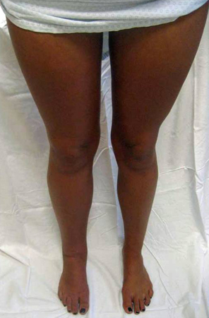 [Figure caption and citation for the preceding image starts]: Secondary lymphoedema: left lower extremity lymphoedema following radiotherapy and lymphadenectomy for Hodgkin's lymphomaFrom the collection of Dr Arin K. Greene [Citation ends].
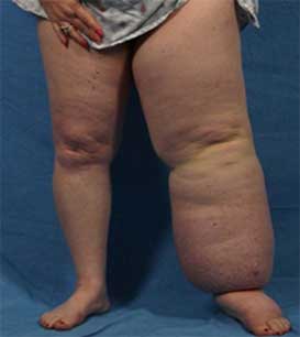
Investigations
When history and physical examination are equivocal, imaging can facilitate the diagnosis, and may also help with treatment planning and monitoring of disease progression. Lymphoscintigraphy, which has been used for >50 years, visualises a radiolabelled marker as it moves through the lymphatic system.[2]Brouillard P, Witte MH, Erickson RP, et al. Primary lymphoedema. Nat Rev Dis Primers. 2021 Oct 21;7(1):77.
http://www.ncbi.nlm.nih.gov/pubmed/34675250?tool=bestpractice.com
[47]Executive Committee of the International Society of Lymphology. The diagnosis and treatment of peripheral lymphedema: 2020 consensus document of the International Society of Lymphology. Lymphology. 2020;53(1):3-19.
https://journals.uair.arizona.edu/index.php/lymph/article/download/23775/22411
http://www.ncbi.nlm.nih.gov/pubmed/32521126?tool=bestpractice.com
Lymphoscintigraphy has a 96% sensitivity and 100% specificity for the disease.[56]Hassanein AH, Maclellan RA, Grant FD, et al. Diagnostic accuracy of lymphoscintigraphy for lymphedema and analysis of false-negative tests. Plast Reconstr Surg Glob Open. 2017 Jul 12;5(7):e1396.
https://journals.lww.com/prsgo/fulltext/2017/07000/Diagnostic_Accuracy_of_Lymphoscintigraphy_for.16.aspx
http://www.ncbi.nlm.nih.gov/pubmed/28831342?tool=bestpractice.com
Lymphangiography involves injecting lymphatic channels with radio-opaque contrast dye and gives a quantitative analysis of lymph flow; however, it is rarely used because of its associated morbidity.[18]Slavin SA, Greene AK, Borud LJ. Lymphedema. In: Weinzweig J, ed. Plastic surgery secrets plus. 2nd ed. Philadelphia, PA: Mosby; 2009. However, it may be helpful in determining the location of a specific anatomical obstruction for preoperative planning of a bypass procedure.[18]Slavin SA, Greene AK, Borud LJ. Lymphedema. In: Weinzweig J, ed. Plastic surgery secrets plus. 2nd ed. Philadelphia, PA: Mosby; 2009.
Magnetic resonance imaging (MRI) techniques encompassing MR lymphography (MRL) and MR angiography (MRA) both with and without contrast (peripheral or intranodal) are being increasingly utilised at centres globally, and provide high resolution imaging with no ionising radiation.[47]Executive Committee of the International Society of Lymphology. The diagnosis and treatment of peripheral lymphedema: 2020 consensus document of the International Society of Lymphology. Lymphology. 2020;53(1):3-19.
https://journals.uair.arizona.edu/index.php/lymph/article/download/23775/22411
http://www.ncbi.nlm.nih.gov/pubmed/32521126?tool=bestpractice.com
[57]Notohamiprodjo M, Weiss M, Baumeister RG, et al. MR lymphangiography at 3.0 T: correlation with lymphoscintigraphy. Radiology. 2012 Jul;264(1):78-87.
http://www.ncbi.nlm.nih.gov/pubmed/22523325?tool=bestpractice.com
[58]Pieper CC, Feisst A, Schild HH. Contrast-enhanced interstitial transpedal MR lymphangiography for thoracic chylous effusions. Radiology. 2020 May;295(2):458-66.
http://www.ncbi.nlm.nih.gov/pubmed/32208098?tool=bestpractice.com
Computed tomography (CT) may also be used, although it exposes the patient to ionising radiation, and has it has demonstrated lower levels of diagnostic and prognostic accuracy.[59]Kim SY, Bae H, Ji HM. Computed tomography as an objective measurement tool for secondary lymphedema treated with extracorporeal shock wave therapy. Ann Rehabil Med. 2015 Jun;39(3):488-93.
https://e-arm.org/journal/view.php?doi=10.5535/arm.2015.39.3.488
http://www.ncbi.nlm.nih.gov/pubmed/26161357?tool=bestpractice.com
Lymphoscintigraphy combined with single photon emission CT (LAS-SPECT-CT) can produce higher resolution images and improve spatial localisation.[47]Executive Committee of the International Society of Lymphology. The diagnosis and treatment of peripheral lymphedema: 2020 consensus document of the International Society of Lymphology. Lymphology. 2020;53(1):3-19.
https://journals.uair.arizona.edu/index.php/lymph/article/download/23775/22411
http://www.ncbi.nlm.nih.gov/pubmed/32521126?tool=bestpractice.com
If history arouses suspicion of filariasis then blood smears for detection of microfilariae are indicated. Genetic testing may be ordered at specialist centres for patients with primary lymphoedema.[47]Executive Committee of the International Society of Lymphology. The diagnosis and treatment of peripheral lymphedema: 2020 consensus document of the International Society of Lymphology. Lymphology. 2020;53(1):3-19.
https://journals.uair.arizona.edu/index.php/lymph/article/download/23775/22411
http://www.ncbi.nlm.nih.gov/pubmed/32521126?tool=bestpractice.com
[Figure caption and citation for the preceding image starts]: Lymphoscintigraphy showing dermal back-flow and absent uptake of radiolabelled colloid in lymph nodes of left lower extremity consistent with lymphoedemaFrom the collection of Dr Arin K. Greene [Citation ends].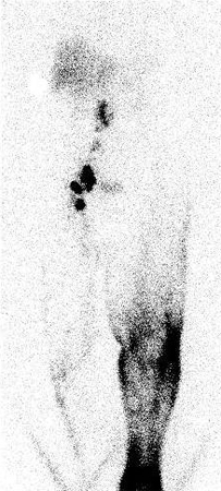
Typically, next-generation sequencing techniques are used to screen blood-derived DNA using gene panels. Whole exome sequencing is another increasingly used option.[2]Brouillard P, Witte MH, Erickson RP, et al. Primary lymphoedema. Nat Rev Dis Primers. 2021 Oct 21;7(1):77.
http://www.ncbi.nlm.nih.gov/pubmed/34675250?tool=bestpractice.com