Images and videos
Images

Assessment of peripheral oedema
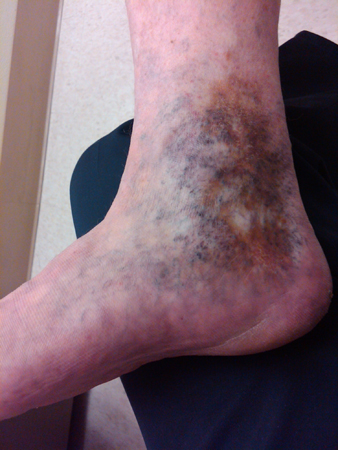
Atrophie blanche and haemosiderin deposition in a patient with chronic venous insufficiency
From the collection of Dr Joseph L. Mills; used with permission
See this image in context in the following section/s:

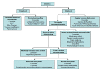
Assessment of peripheral oedema
Approach to the patient with peripheral oedema. DVT, deep vein thrombosis; HTN, hypertension
Created by Ethan Cumbler, MD
See this image in context in the following section/s:
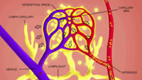
Assessment of peripheral oedema
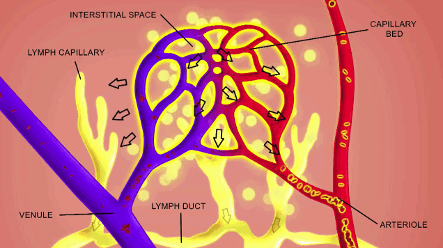
Development of oedema: an imbalance between the filtration of fluid from the bloodstream and drainage via the lymph system
Created by BMJ Knowledge Centre
See this image in context in the following section/s:
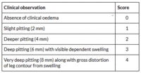
Assessment of peripheral oedema
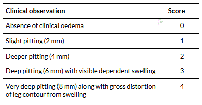
A clinical assessment scoring system
Created by BMJ Knowledge Centre based on Brodovicz KG, McNaughton K, Uemura N, et al. Reliability and feasibility of methods to quantitatively assess peripheral edema. Clin Med Res. 2009 Jun;7(1-2):21-31.
See this image in context in the following section/s:

Assessment of peripheral oedema
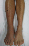
Deep vein thrombosis (DVT) of the right leg with associated swelling and redness
From the collection of James Heilman, MD.
See this image in context in the following section/s:
Videos
 Venepuncture and phlebotomy animated demonstration
How to take a venous blood sample from the antecubital fossa using a vacuum needle.
 How to perform an ECG animated demonstration
How to record an ECG. Demonstrates placement of chest and limb electrodes.
Use of this content is subject to our disclaimer