Recommendations
Urgent
Suspect a tension pneumothorax if there is sudden onset of:[8][40]
Cardiopulmonary deterioration
Hypotension; this suggests imminent cardiac arrest
Respiratory distress
Low oxygen saturations
Tachycardia
Shock
Loss of consciousness
Severe chest pain
Sweating.
Examine for ipsilateral reduced breath sounds, reduced chest expansion, hyper-resonance on percussion, and tracheal shift to the contralateral side.
Put out an immediate cardiac arrest call and give high-flow oxygen. Perform emergency needle decompression; do not wait for imaging to confirm the diagnosis. Insert a chest drain following this.[17][41][42] See Management Recommendations.
Maintain a high level of suspicion for a tension pneumothorax in a patient with any of the following risk factors:[43]
Ventilated patients
Following trauma (especially penetrating chest wounds) or cardiopulmonary resuscitation
Lung disease, especially acute presentations of asthma and bullous COPD, or long-standing lung disease such as cystic fibrosis, bronchiectasis, fibrotic lung disease, or lung cancer
Blocked chest drain
Patients receiving non-invasive ventilation (NIV)
Other (e.g., hyperbaric oxygen treatment).
Key Recommendations
Suspect a non-tension pneumothorax in a stable patient who has sudden onset of chest pain, dyspnoea, or cough, especially if they have risk factors such as smoking or underlying lung disease.[8][44]
Symptoms tend to be more severe in secondary spontaneous pneumothorax and may be minimal or absent in primary spontaneous pneumothorax.[8][45]
Signs on examination include:
Use erect postero-anterior (PA) chest x-ray as the first-line investigation to definitively diagnose a pneumothorax in a stable patient who can sit upright.[17]
Order a CT chest if the diagnosis is uncertain on chest x-ray and the patient remains symptomatic, or in stable patients with significant chest trauma.[17][47] If the patient is stable, discuss with a radiologist.
If pneumothorax is diagnosed, measure the visible rim between the lung margin and the chest wall at the level of the hilum on imaging to help guide further management. The British Thoracic Society recommends that size of pneumothorax is no longer an indication for invasive management; however, it does dictate the safety of conducting an intervention.[17]
Tension pneumothorax is a life-threatening emergency that needs urgent identification and treatment with decompression and high-flow oxygen; do not wait for imaging to confirm the diagnosis.[17] See Management Recommendations.
Signs are typically sudden in onset and include:[8]
Cardiopulmonary deterioration
Hypotension; this suggests imminent cardiac arrest
Respiratory distress
Low oxygen saturations
Tachycardia
Shock
See our topic Shock
Loss of consciousness
Severe chest pain
Sweating.
Practical tip
Tension pneumothorax is extremely rare in a primary spontaneous pneumothorax (PSP).[8] However, significant breathlessness in a patient with a small PSP may indicate the development of a tension pneumothorax.[49]
Other concerning symptoms in a patient with a small PSP that may indicate the development of a tension pneumothorax are chest pain, feeling of dread or panic, excessive sweating, agitation, and confusion.[17][42]
General features of a non-tension pneumothorax also tend to be sudden in onset and include:
Chest pain
Dyspnoea
More prominent in secondary spontaneous pneumothorax (SSP)[8]
Cough
Sometimes present in pneumothorax ex vacuo (commonly known as ‘trapped lung’).[44]
Symptoms of SSP are generally more severe than symptoms of PSP due to coexistent lung disease, and may be present even with relatively small pneumothoraces. Symptoms may be very mild or absent in PSP.[8][45]
Practical tip
Symptoms usually improve following presentation of PSP. If symptoms worsen, consider the development of complications such as tension pneumothorax or haemopneumothorax, or an alternative cause of symptoms.[8]
Look for tracheal shift to the contralateral side, which indicates a tension pneumothorax, although examination can be challenging in this setting.[40]
Look for other general signs of a pneumothorax:
Ipsilateral reduced breath sounds[8]
This is the most common sign in tension pneumothorax
Ipsilateral hyperinflation of the hemithorax with hyper-resonance on percussion[8]
However, hyperinflation may be difficult to detect clinically in a tension pneumothorax, and is not present in a pneumothorax ex vacuo
Hypoxia
Evidence of penetrating trauma or rib fractures in a traumatic pneumothorax.[43]
Remember there may also be signs of underlying lung disease (e.g., COPD) if the pneumothorax is secondary.
Maintain a high level of suspicion for a tension pneumothorax in a patient with any of the following risk factors:[41][43]
Ventilated patients
Following trauma (especially penetrating chest wounds) or cardiopulmonary resuscitation
Lung disease, especially acute presentations of asthma and bullous COPD, or in long-standing underlying lung disease such as cystic fibrosis, bronchiectasis, fibrotic lung diseases, or lung cancer
Blocked chest drain
Patients receiving non-invasive ventilation (NIV)
Other (e.g., hyperbaric oxygen treatment).
Practical tip
Maintain a high level of suspicion for a tension pneumothorax in patients using ventilators who have a rapid onset of haemodynamic instability or cardiac arrest, particularly if they require increasing peak inspiratory pressures.[43]
Consider other general risk factors for pneumothorax, including:
Smoking[8]
This is the most important risk factor; men who smoke increase their risk of a first pneumothorax 22-fold and women 9-fold compared with non-smokers[7]
Family history of pneumothorax[9]
Tall and slender body build[8]
Male sex[8]
Young age[8]
However, secondary spontaneous pneumothorax is more common in people aged >55 years
Presence of underlying lung disease such as:
COPD
Severe asthma
Tuberculosis
Pneumocystis jirovecii infection
Cystic fibrosis
Structural abnormalities (e.g., Marfan syndrome, Ehlers-Danlos syndrome)[10]
Recent invasive medical procedures (e.g., drainage of pleural effusion or CT-guided lung biopsy)[13][14]
Trauma[12]
Homocystinuria[9]
Menstruation
Use imaging to definitively diagnose a pneumothorax and to measure the size of the pneumothorax. However, clinical evaluation is more important than the size of the pneumothorax in determining further management.[17]
Chest x-ray (always order)
Use chest x-ray as the first-line investigation in stable patients who can sit upright to definitively diagnose pneumothorax.[17]
[Figure caption and citation for the preceding image starts]: Anterior-posterior chest x-ray demonstrating a right pneumothoraxFrom the collection of Dr Ryland P. Byrd [Citation ends].
Practical tip
Potential mimics of a pneumothorax on chest x-ray are:[51]
Bullous lung disease
Medial border of the scapula
Outline of the oxygen reservoir bag or associated tubing
Clothing
Bedsheets
Companion shadows (visible subcostal groove usually at ribs 1 and 2)
Skin folds
Post-pleurectomy scarring/suture material.
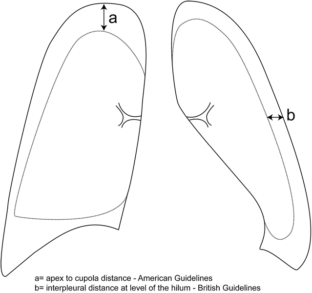
If pneumothorax is confirmed on imaging, measure the visible rim between the lung margin and the chest wall at the level of the hilum. This can be done using chest x-ray but is most accurately measured using CT. Although the size of a pneumothorax is no longer an indication for invasive management, it does dictate the safety of conducting an intervention. Whether a pneumothorax is of sufficient size to intervene depends on clinical context but, in general, is usually ≥2 cm laterally or apically on chest x-ray, or any size on CT scan that can be safely accessed with radiological support:[17]
[Figure caption and citation for the preceding image starts]: UK guidelines advise using the level of the hilum to measure the size of a pneumothorax. However, other countries may use other methods; for example US guidelines use the distance from the lung apex to the cupola, but this method would tend to overestimate the volume of a localised apical pneumothorax.Copyright © BMJ Publishing Group Ltd and British Thoracic Society. All rights reserved. [Citation ends]. In the UK, the level of the hilum is usually used to measure the size of a pneumothorax. However, other countries may use other methods; for example US guidelines use the distance from the lung apex to the cupola, although this method would tend to overestimate the volume of a localised apical pneumothorax.
Note that the size of pneumothorax on imaging does not correlate well with the clinical presentation of the patient.
A 2 cm visible rim corresponds to a pneumothorax of about 50% (complete collapse of the lung is a 100% pneumothorax).[43]
Blood tests (always order)
Order a full blood count and clotting screen.
Correct clotting abnormalities (INR ≥1.5 or platelets ≤50 x 109/L) before inserting a chest drain in patients who are not critically unwell.[52][53]
Chest ultrasound (consider ordering)
Chest ultrasound is increasingly used to detect pneumothorax, especially for patients who are immobilised following trauma, when an erect PA chest x-ray cannot be obtained. It requires specialist expertise.[54][55][56]
Evidence: Ultrasound for diagnosis of pneumothorax
Ultrasound may be helpful for trauma patients who are unable to undergo erect chest x-ray.
One Cochrane systematic review (search date April 2020) compared chest ultrasound by frontline non‐radiologist physicians with portable supine anterior-posterior chest x-ray for the diagnosis of pneumothorax in trauma patients in the emergency department. CT of the chest or tube thoracostomy were used as the reference standard.[56]
It found 13 prospective, paired accuracy studies of which 9 (410 patients with traumatic pneumothorax) used patients, as opposed to lung field, as the unit of analysis.
All studies were at high or unclear risk of bias in at least one domain.
Chest ultrasound was more sensitive than chest x-ray (ultrasound 0.91, 95% CI 0.85 to 0.94; chest x-ray 0.47, 95% CI 0.31 to 0.63). Both had high specificity with no significant difference between chest ultrasound and x-ray for specificity.
This means that (assuming a prevalence of 30%) there would be around 5 times the numbers of missed traumatic pneumothorax (false negatives) with chest x-ray compared with ultrasound, whilst both have very low false positive rates.
Further analyses found these results were independent of the type of trauma (blunt or penetrating), ultrasound operator, or type of ultrasound probe used.
CT chest (consider ordering)
Order a CT chest if the diagnosis is uncertain on chest x-ray and the patient remains symptomatic, or in stable patients with significant chest trauma. If the patient is stable discuss this with a radiologist.[47] CT guidance may be required in some situations, including loculated pneumothorax with tethered lung, the presence of bullae, or posteriorly loculated pleural fluid collections, where sonographic views are not optimal.[52]
CT chest is considered the gold standard for accurate assessment of the size of the pneumothorax.
CT chest can also be used to:[57]
Differentiate pneumothorax from bullous lung disease
Identify underlying lung disease in patients with secondary spontaneous pneumothorax.
Arterial blood gas (consider ordering)
Consider an arterial blood gas (ABG) if oxygen saturations are ≤92% on room air. It may help rule out other differential diagnoses but is not usually necessary.
Escalate to a senior colleague if there is acute respiratory acidosis.
Respiratory alkalosis is the most common finding.[58]
Use of this content is subject to our disclaimer