Images and videos
Images


Assessment of red eye
Corneal abrasion seen with fluorescein stain
Private collection - courtesy of Mr Hugh Harris
See this image in context in the following section/s:

Assessment of red eye
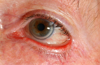
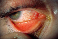
Ectropion
Private collection - courtesy of Mr Hugh Harris
See this image in context in the following section/s:
Assessment of red eye
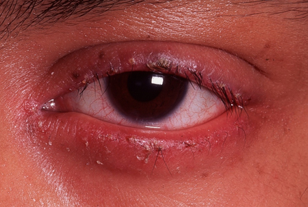
Blepharitis
Private collection - courtesy of Mr Hugh Harris
See this image in context in the following section/s:

Assessment of red eye
Corneal foreign body
Private collection - courtesy of Mr Hugh Harris
See this image in context in the following section/s:

Assessment of red eye
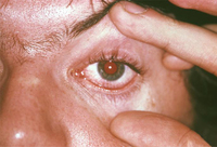
A patient with left herpes zoster ophthalmicus affecting the forehead and side of the nose (positive Hutchinson’s sign; yellow arrows). The crusted skin rashes follow the V1 dermatomal distribution and do not cross the vertical midline
Image used with permission from BMJ 2019;364:k5234
See this image in context in the following section/s:

Assessment of red eye
Viral conjunctivitis
Private collection - courtesy of Mr Hugh Harris
See this image in context in the following section/s:
Assessment of red eye
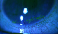
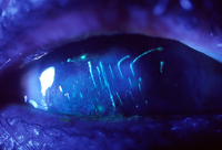
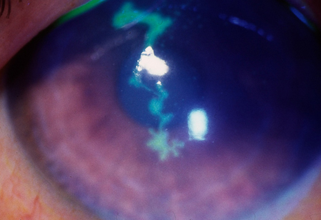
Dendritic ulcer seen with fluorescein stain
Private collection - courtesy of Mr Hugh Harris
See this image in context in the following section/s:

Assessment of red eye
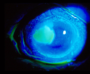
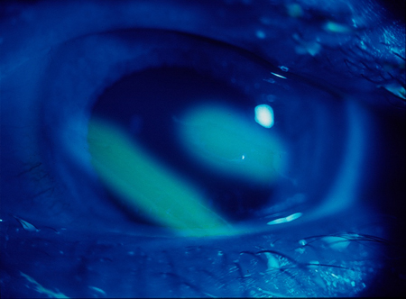
Corneal ulcer seen with fluorescein stain
Private collection - courtesy of Mr Hugh Harris
See this image in context in the following section/s:

Assessment of red eye
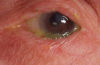
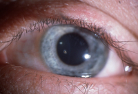
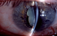
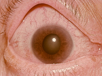
Angle-closure glaucoma: central corneal oedema with an oval-shaped mid-dilated pupil.
Private collection - courtesy of Mr Hugh Harris
See this image in context in the following section/s:

Assessment of red eye
Subtarsal foreign body: vertical corneal abrasions seen with fluorescein stain
Private collection - courtesy of Mr Hugh Harris
See this image in context in the following section/s:
Assessment of red eye
Gonorrhoeal conjunctivitis: resulted in partial blindness
CDC Image Library
See this image in context in the following section/s:
Assessment of red eye
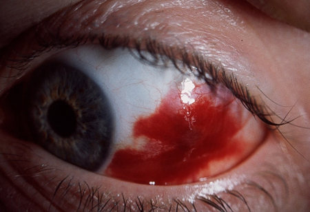
Subconjunctival haemorrhage
Private collection - courtesy of Mr Hugh Harris
See this image in context in the following section/s:
Assessment of red eye
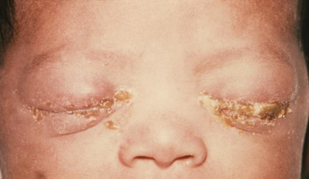
A newborn with gonococcal ophthalmia neonatorum caused by a maternally transmitted gonococcal infection
US Centers for Disease Control and Prevention/ J. Pledger
See this image in context in the following section/s:
Assessment of red eye
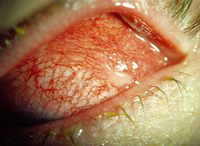
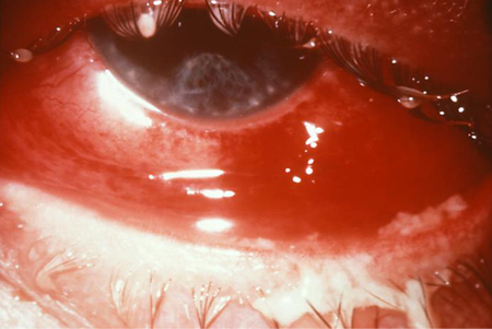
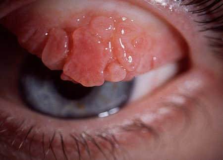
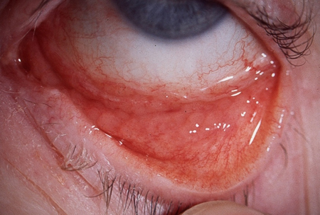
Allergic (vernal) keratoconjunctivitis
Private collection - courtesy of Mr Hugh Harris
See this image in context in the following section/s:
Assessment of red eye
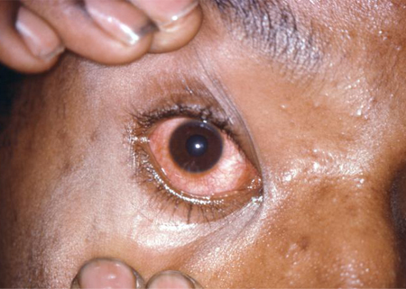
Gonococcal conjunctivitis
CDC Image Library/Joe Miller
See this image in context in the following section/s:

Assessment of red eye
Conjunctivitis: consequence of reactive arthritis
CDC Image Library/Joe Miller
See this image in context in the following section/s:

Assessment of red eye
Dry eye (stained with fluorescein)
From the personal collection of Dr Jonathan Smith; used with permission
See this image in context in the following section/s:

Assessment of red eye
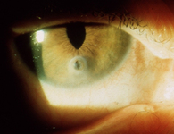
Anterior uveitis with posterior synechiae
Private collection - courtesy of Mr Hugh Harris
See this image in context in the following section/s:

Assessment of red eye
Bacterial conjunctivitis
Private collection - courtesy of Mr Hugh Harris
See this image in context in the following section/s:
Assessment of red eye
Chlamydial conjunctivitis
Private collection - courtesy of Mr Hugh Harris
See this image in context in the following section/s:

Assessment of red eye
Episcleritis
Private collection - courtesy of Mr Hugh Harris
See this image in context in the following section/s:

Assessment of red eye
Trichiasis
Private collection - courtesy of Mr Hugh Harris
See this image in context in the following section/s:

Assessment of red eye
Entropion
Private collection - courtesy of Mr Hugh Harris
See this image in context in the following section/s:

Assessment of red eye
Penetrating corneal injury with iris prolapse
Private collection - courtesy of Mr Hugh Harris
See this image in context in the following section/s:
Assessment of red eye
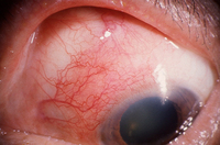
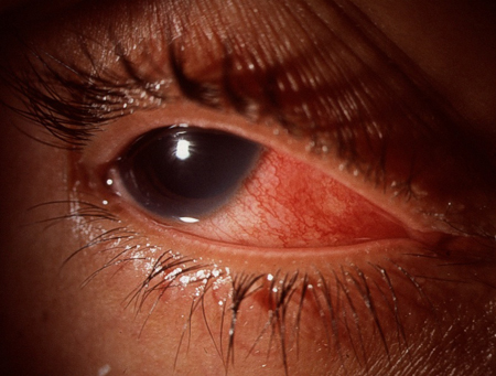
Scleritis
Private collection - courtesy of Mr Hugh Harris
See this image in context in the following section/s:
Use of this content is subject to our disclaimer