Images and videos
Images

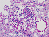


IgA nephropathy
Endocapillary hypercellularity in IgA nephropathy (Periodic acid-Schiff stain, x400)
Courtesy of Drs Hwei Yee Lee, Cristine Ding, and Yong Howe Ho (Tan Tock Seng Hospital, Singapore)
See this image in context in the following section/s:

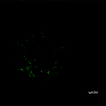
IgA nephropathy
Immunofluorescence staining for IgA showing strong granular staining in the mesangium globally (anti-IgA immunofluorescence, x200)
Courtesy of Drs Hwei Yee Lee, Cristine Ding, and Yong Howe Ho (Tan Tock Seng Hospital, Singapore)
See this image in context in the following section/s:
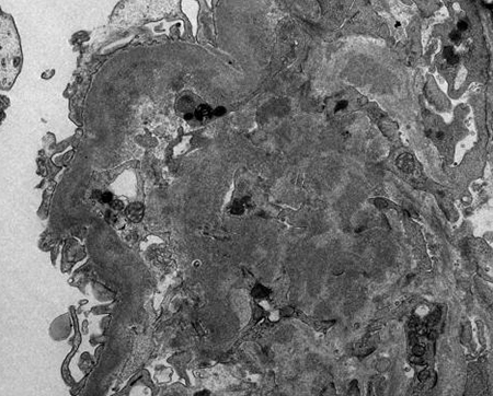
IgA nephropathy
Electron dense deposits in mesangial and paramesangial regions (electron micrograph, x1000)
Courtesy of Drs Hwei Yee Lee, Cristine Ding, and Yong Howe Ho (Tan Tock Seng Hospital, Singapore)
See this image in context in the following section/s:

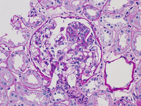
IgA nephropathy
Segmental glomerulosclerosis in IgA nephropathy (Periodic acid-Schiff stain, x400)
Courtesy of Drs Hwei Yee Lee, Cristine Ding, and Yong Howe Ho (Tan Tock Seng Hospital, Singapore)
See this image in context in the following section/s:
IgA nephropathy
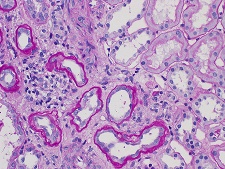
Tubular atrophy and interstitial fibrosis in IgA nephropathy (the atrophied tubules are seen on the left of the field, and normal tubules are seen on the right) (Periodic acid-Schiff stain, x400)
Courtesy of Drs Hwei Yee Lee, Cristine Ding, and Yong Howe Ho (Tan Tock Seng Hospital, Singapore)
See this image in context in the following section/s:
IgA nephropathy
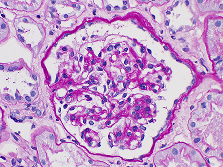
Mesangial hypercellularity in IgA nephropathy (Periodic acid-Schiff stain, x600)
Courtesy of Drs Hwei Yee Lee, Cristine Ding, and Yong Howe Ho (Tan Tock Seng Hospital, Singapore)
See this image in context in the following section/s:
Use of this content is subject to our disclaimer