Approach
Symptoms of acute infectious laryngitis may range from very subtle features to high-grade fever with airway compromise. Subtle features may include short-lived mild hoarseness and upper respiratory infection symptoms. The clinical presentation of laryngitis depends on:
Causative pathogen
Amount of tissue oedema
Region of larynx primarily involved
Age and comorbidities.
Evaluation of the airway is important as an initial step. Further evaluation then follows.
Urgent considerations
Upon presentation of acute laryngitis, the first system assessed should be the airway. If there is respiratory distress, the patient should be assessed in a controlled environment with the facility to perform safe intubation. Emergency tracheotomy may be required if, through swelling, a normal intubation is not possible.
Children presenting with symptoms and signs of epiglottitis (e.g., high fever, sore throat, toxic appearance, drooling, tripod positioning, difficulty breathing, and irritability) should be examined in a controlled setting, such as the operating room. Intubation is performed if there is any doubt about the airway. See Epiglottitis.
If the patient is an adult, flexible laryngoscopy may be performed, depending on the level of distress. If the patient is in severe respiratory distress, or if supraglottitis is suspected, then flexible laryngoscopy may trigger laryngospasm and airway demise. In these patients laryngeal examination should be performed only by an otolaryngologist, and preferably in the operating room where a surgical airway can be secured if needed. Any manipulation of the supraglottic area should be avoided. If necessary, intubation can be performed during flexible laryngoscopy with direct visualisation.[19]
History
Once the airway is assessed and, if necessary, secured, the remainder of the history and examination can be performed. A thorough history should include information about voice, breathing, and swallowing patterns. Concomitant systemic problems, such as allergies, exposures, immune deficiencies, and systemic illnesses, are considered. History of intubations, radiation exposures, and neck surgery should be taken, as should a smoking history.
Knowledge of recent travel to areas where diphtheria or tuberculosis are endemic, or of contact with people with infectious symptoms, may aid in diagnosis.[12] Other risk factors for acute infectious laryngitis include incomplete or absent Haemophilus influenzae type B (Hib) or diphtheria vaccination. Laryngeal candidiasis is more common in patients using inhaled corticosteroids or prolonged courses of antibiotics, and in those who are immune compromised.[12]
Patients with acute infectious laryngitis can present with symptoms ranging from very subtle features to high-grade fever and airway compromise. There is often a preceding upper respiratory tract infection with sore throat, fever, cough, and rhinitis. This is followed by odynophagia, dysphagia, and hoarseness. Fatigue and malaise can occur. Laryngeal oedema can lead to dyspnoea.
A challenge for the clinician is to decide which patients may have bacterial infection and require specific antibiotic treatment, because presentation of viral and bacterial laryngitis may be similar.[14] Viral laryngitis is common, and symptoms generally arise over a period of <7 days. Evidence of a bacterial infection elsewhere (e.g., pneumonia, streptococcal pharyngitis) supports a bacterial aetiology.[4] Diphtheria is uncommon in the US and has a prodrome of several days, with hoarseness progressing to airway compromise. See Diphtheria.
Chronic laryngitis is defined as throat inflammation of at least 3-week duration that encompasses a broad range of inflammatory, infectious, and autoimmune conditions resulting in alteration of phonation, breathing, and swallowing. Symptoms include dysphonia, throat pain, globus sensation, frequent throat clearing, cough, and dysphagia.[1] The symptoms of chronic laryngitis due to TB are prolonged (>3 weeks) and patients typically complain of dysphonia, but may also experience odynophagia, dysphagia, coughing, and rarely dyspnoea.[1]
Symptoms generally mimic symptoms of laryngeal malignancy, which needs to be ruled out. Patients may have symptoms of cough and weight loss, but they are usually referred to an otolaryngologist due to persistent hoarseness.
Patients with traumatic laryngitis will present with hoarseness that has been going on for a prolonged duration and generally have a history of heavy vocal use. These patients tend to be professional voice users such as teachers, lawyers, people in sales, or singers. The hoarseness is usually worse with increased voice use, therefore, they have more complaints towards the end of the day and are better in the morning. If they do voice rest, they tend to have improved voice quality. If there is an acute trauma, they can present with sudden onset loss of voice, which could be a sign of a vocal fold haemorrhage.
Physical examination
Generally, an adult with acute laryngitis will not be toxic in the absence of acute epiglottitis or diphtheria. Patients may have hyperaemia of the oropharynx and possibly enlarged tonsils. There may be post-nasal drip on oropharyngeal examination. Exudative tonsillopharyngitis, anterior cervical lymphadenitis, and fever are highly suggestive of a bacterial origin.[14]
A patient with diphtheria can present with a sore throat, difficulty swallowing, malaise or be in acute respiratory distress. Oropharyngeal examination can reveal white-grey exudates, which may extend to the soft palate and vallecula. These pseudomembranes may also be found covering the laryngeal structures, leading to airway compromise. Exudates are firmly adherent to the underlying mucosa, which bleed when the exudate is removed. There is cervical lymphadenopathy, profound malaise, and stridor. The diphtheria toxin also causes cardiomyopathy and neuropathies.[20] Paralysis of the vocal folds or palate can be seen. Early diagnosis is imperative. See Diphtheria. [Figure caption and citation for the preceding image starts]: Young boy presenting with acute diphtheria infectionImage courtesy of CDC [Citation ends].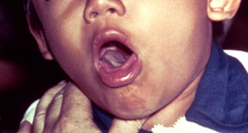
Head and neck examination is usually normal in vocal trauma.
Patients with chronic laryngitis secondary to reflux may demonstrate laryngeal oedema, pseudosulcus, hyperaemia, increased mucus, granuloma, or thickening of the posterior interarytenoid tissue.[21]
Diagnostic tests
Laryngitis is a diagnosis of history and examination, rather than laboratory testing. A thorough examination includes laryngoscopy. This is performed if the patient presents initially to an otolaryngology specialist, but most primary care physicians are not experienced in the technique and diagnose most cases of viral laryngitis clinically. Some primary care physicians may use mirror indirect laryngoscopy, depending on experience. Laryngoscopy shows oedema and erythema of the laryngeal structures, especially the true vocal folds. Thick, copious, white-yellow secretions are also seen in the glottis. If indirect laryngoscopy cannot be performed, the patient may be referred to an otolaryngology specialist. Indications for referral to an otolaryngologist include:
Uncertain diagnosis.
Persistent hoarseness (lasting longer than 2-3 weeks) with failed treatment. It is important to refer these patients rather than treat them with further courses of antibiotics if symptoms do not improve or resolve within 4 weeks, or earlier if a serious underlying cause is suspected.[22]
Ill patients with suspected airway compromise. These patients are referred to hospital for urgent management and assessment.
Patients whose profession relies on their voice.
Videostroboscopy allows for simultaneous evaluation of voice quality, laryngeal anatomy, and vocal fold vibratory function.[23] Guidelines on dysphonia encourage the use of videostroboscopic examination when the voice symptoms are out of proportion to the indirect laryngoscopy.[22] Videostroboscopy can reveal vocal fold sulcus or vibratory pathologies such as stiffness, or help differentiate between benign vocal lesions.[22]
In case of suspected bacterial origin, oropharyngeal cultures and full blood countcan be obtained, as well as a rapid antigen detection test. If diphtheria is suspected, cultures of nose and throat swabs are obtained and Loeffler or Tindale selective media used. Definitive diagnosis can also be made by the demonstration of toxin production by immunoprecipitation or polymerase chain reaction.
The work-up for patients with chronic laryngitis suspected to be due to tuberculosis includes a chest x-ray, sputum cultures, and sputum smear for the detection of acid-fast bacilli. See Pulmonary Tuberculosis. Indirect laryngoscopy generally reveals exophytic or nodular lesions. Most commonly, the posterior glottis is involved, but the lesions can be seen anywhere in the larynx. Because the laryngeal lesions look similar to carcinoma of the larynx, a direct laryngoscopy should be performed, and biopsies should be obtained.[22] This procedure is usually performed under general anaesthesia by an otolaryngologist.
In vocal strain, history and examination to exclude other causes is usually sufficient to make the diagnosis. However, other aetiologies may exist in heavy voice users (including laryngeal malignancy); therefore, any hoarseness that does not improve or resolve within 4 weeks should be evaluated by an otolaryngologist with a laryngoscopy. If a serious underlying cause is suspected, the patient should be referred irrespective of duration.[22]
Use of this content is subject to our disclaimer