Images and videos
Images

Assessment of dysmenorrhoea
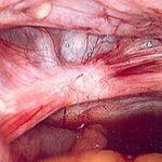
Laparoscopic image of endometriotic nodule
From the collection of Dr Jonathon Solnik; used with permission
See this image in context in the following section/s:
Assessment of dysmenorrhoea
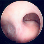
Multiple polyps are identified on hysteroscopic examination of the uterine cavity in this patient with persistent vaginal spotting
From the collection of Dr M.F. Mitwally and Dr R.J. Fischer; used with permission
See this image in context in the following section/s:

Assessment of dysmenorrhoea
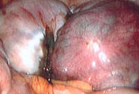
Laparoscopic image of ovarian endometrioma
From the collection of Dr Jonathon Solnik; used with permission
See this image in context in the following section/s:

Assessment of dysmenorrhoea
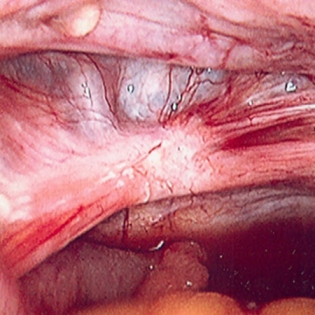
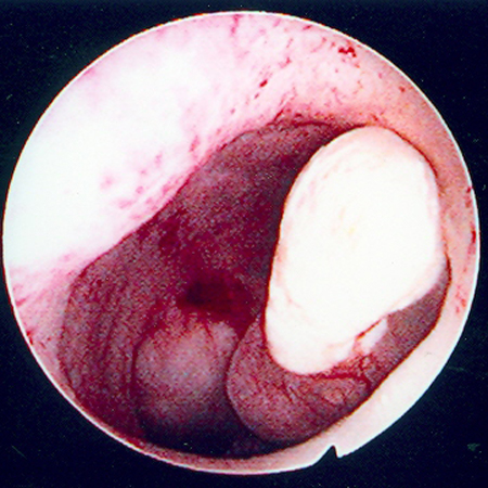
Hysteroscopic image of a large pedunculated submucous uterine fibroid
From the collection of Dr M.F. Mitwally and Dr R.J. Fischer; used with permission
See this image in context in the following section/s:
Use of this content is subject to our disclaimer