Investigations
1st investigations to order
non-contrast CT head
Test
Order an urgent non-contrast CT head, to be done as soon as possible, for all patients presenting with acute sudden, severe headache (thunderclap headache) or other signs and symptoms that suggest SAH.[38][39][43][51]
Ideally, the CT scan should be within 6 hours of symptom onset.[38]
There is good evidence showing that non-contrast CT head scans carried out within 6 hours of symptom onset are highly accurate and can be used to rule out a diagnosis of SAH. CT head scans done more than 6 hours after symptom onset are less accurate.[38][51]
Modern, third-generation scanners detect SAH in 93% of cases if done in the first 24 hours after the bleed, and in almost 100% of cases when performed within 6 hours of onset of headache and interpreted by an experienced radiologist.[76][77][78][79] The sensitivity of CT in detecting SAH declines after the first 24 hours to 68% on day 3 and 58% on day 5.[8][9]
CT is the standard diagnostic test for SAH.
Practical tip
Some patients with SAH present with vomiting and/or are agitated and require anaesthesia to be scanned. Call the anaesthetist as soon as possible to not delay diagnosis in these patients.
Order thin cuts (3-5 mm) to avoid missing small, thin collections of blood.
State on the CT request that SAH is being considered so that the appropriate thin slices are performed and the radiologist knows what they are looking for.
Practical tip
Look for hydrocephalus on the CT. This may explain decreased level of consciousness (low or falling Glasgow Coma Scale score).
Once the diagnosis of SAH is confirmed, urgently discuss with a specialist neurosurgical centre the need for transfer of care of the patient to the specialist centre.[38]
Do not use a SAH severity score in isolation to determine the need for, or timing of, transfer of care to a specialist neurosurgical centre.[38][52]
[Figure caption and citation for the preceding image starts]: CT brain showing subarachnoid haemorrhage from a ruptured posterior cerebral artery aneurysm (1 of 2)Courtesy of Dr Salah Keyrouz; used with permission [Citation ends].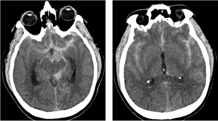 [Figure caption and citation for the preceding image starts]: CT brain showing subarachnoid haemorrhage from a ruptured posterior cerebral artery aneurysm (2 of 2)Courtesy of Dr Salah Keyrouz; used with permission [Citation ends].
Result
hyperdense areas in the subarachnoid space/basal cisterns
MRI
Test
While CT is the preferred imaging modality for confirming SAH, the European Stroke Organization states that MRI with multiple sequences is equally suitable for diagnosis within the first 24 hours.[43]
Once the diagnosis of SAH is confirmed, urgently discuss with a specialist neurosurgical centre the need for transfer of care of the patient to the specialist centre.[38]
Result
increased signal intensity in the subarachnoid space on fluid-attenuated inversion recovery (FLAIR) images
FBC
Test
Leukocytosis following SAH is an independent risk factor for cerebral vasospasm.[80]
Result
may show leukocytosis
serum electrolytes
Test
Hyponatraemia is the most common electrolyte abnormality in SAH. It is usually associated with syndrome of inappropriate antidiuretic hormone secretion (SIADH) although another common cause is cerebral salt-wasting syndrome.[12]
Result
may show moderate to severe hyponatraemia (sodium levels <131 mmol/L [<131 mEq/L])[117]
clotting profile
Test
Baseline coagulation disturbances are common in SAH and need to be treated. Coagulopathy may be present.
Result
may show elevated INR, prolonged PTT
troponin I
serum glucose
Test
Hyperglycaemia develops in one third of SAH patients and is a feature of any acute brain injury.
It is associated with poor clinical condition on admission and independently associated with poor outcome.[43]
Result
may be elevated
ECG
Test
Request an ECG in all patients. Half of patients have an abnormal ECG on admission.[86] Common cardiac findings in SAH include:
Consult immediately with a cardiologist any ECG changes.
Practical tip
Patients with SAH may have ECG changes that mimic acute coronary syndrome and ST-elevation myocardial infarction. Seek specialist advice when deciding whether to perform a CT head scan before or after coronary angiography. Use your clinical judgement alongside specialist input to weigh up the likelihood of the patient having SAH versus acute coronary syndrome.[87]
Aneurysm rupture can lead to cardiac complications, such as left ventricular subendocardial injury and takotsubo cardiomyopathy (even in the absence of acute coronary disease) leading to treatment delays.[43] SAH can also cause cardiac arrest.[88]
Cardiac abnormalities in SAH may be due to massive catecholamine release resulting in overwhelming sympathetic activation.[59][60][61][62] As a result, transient myocardial ischaemia and failure may occur.
[Figure caption and citation for the preceding image starts]: ECG done on admission of a patient with subarachnoid haemorrhage; note peaked, tall T waves (1 of 2)Courtesy of Dr Salah Keyrouz; used with permission [Citation ends].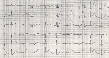
Perform continuous ECG monitoring in all patients at least until occlusion of the aneurysm.[43]
Result
arrhythmias, prolonged QT, ST segment, or T-wave abnormalities (e.g., tall T waves)
Investigations to consider
lumbar puncture (LP)
Test
If a CT head scan performed more than 6 hours after symptom onset is negative or inconclusive, the UK National Institute for Health and Care Excellence (NICE) recommends:[38][51]
To consider an LP
Waiting for at least 12 hours to pass from the onset of symptoms before performing an LP (if LP is indicated)
In the UK, standard practice is to perform an LP 12 hours after the onset of symptoms, or within 14 days if presentation is delayed.[39][89] This is because of the time it takes for red blood cells to lyse and for xanthochromia (elevated bilirubin) to be detected by spectrophotometric analysis of cerebrospinal fluid (CSF).
If a CT head scan performed within 6 hours of symptom onset is reported and documented by a radiologist to show no evidence of SAH, NICE recommends:[38]
Against routinely offering a lumbar puncture (LP)
To consider alternative diagnoses and seek advice from a specialist.
Note, however, that guidelines differ on this point. The European Stroke Organization recommends that lumbar puncture must be performed in any patient with clinically suspected SAH despite negative CT findings. It advises to do this 6-12 hours after symptom onset because blood degradation can take some hours.[43]
Collect 3 tubes of CSF labelled 1 to 3. This is standard practice in the UK.
This helps if the tap has been traumatic (i.e., bleeding from needle insertion when performing LP) as a reduction in the number of red blood cells from tube 1 to tube 3 indicates traumatic tap whereas the number of red blood cells will remain the same if the presence of blood in the CSF is due to SAH.
Exclude SAH if there is a combination of a red blood cell count less than 2000 × 10 6/L and there is no xanthochromia in patients with traumatic lumbar puncture.[90]
Practical tip
Bilirubin degrades in sunlight. Place the sample immediately in an opaque bag (brown bag). This prevents in-vitro degradation of bilirubin to deoxyhaemoglobin, which may lead to false-negative results.[89]
Always use spectrophotometric analysis of CSF as visual inspection for xanthochromia is unreliable.[39][89]
Spectrophotometric analysis of CSF may lead to false-positive results and unnecessary investigation whereas visual inspection for xanthochromia may lead to false-negatives and missed SAH.[89] UK organisations NEQAS (National External Quality Assessment Service) and the Royal College of Physicians recommend always using spectrophotometry analysis, rather than visual inspection.[39][89] However, many countries do not use spectrometry and rely on visual inspection of CSF for reporting xanthochromia. The number of red cells in each sample can also assist in diagnosis.
Diagnose a subarachnoid haemorrhage if the LP sample shows evidence of xanthochromia on spectrophotometry.[38] Consider alternative diagnoses if the LP sample shows no evidence of xanthochromia on spectrophotometry.[38]
Practical tip
Possible reasons for a false-positive (CSF looks xanthochromic) result include:[89][91]
High protein content in CSF
Contamination with iodine used for disinfection
Perimesencephalic haemorrhage
Brain or spinal arteriovenous malformations
Cerebral arterial dissection
Vasculitis
Stroke
Venous sinus thrombosis
Sickle cell disease
Pituitary apoplexy
Substance abuse.
Measure CSF opening pressure in all patients with sudden onset of severe headache. Abnormal CSF pressure may indicate an alternative diagnosis.[46]
If elevated: consider cerebral venous sinus thrombosis or idiopathic intracranial hypertension. See Idiopathic intracranial hypertension.
If reduced: consider low pressure headache.
Once the diagnosis of SAH is confirmed, urgently discuss with a neurosurgical centre the need for transfer of care of the patient to the specialist centre.[38]
Exclude SAH if both CT and LP are negative.[104] This strategy is valid up to 2 weeks from bleed because the sensitivity of LP decreases 2 weeks after the bleed.[89]
Evidence: Lumbar puncture
A lumbar puncture only adds further diagnostic yield in a very-high-probability patient for SAH.
There is good evidence that a normal CT scan performed within 6 hours from headache onset in a person with normal neurological examination effectively excludes SAH.[51][77][78][79] In practice, most patients do not present to hospital within 6 hours from onset of symptoms. Therefore, in these patients, a lumbar puncture (LP) is often carried out if the CT scan is negative or inconclusive. However, LP is an invasive method with reported low specificity, low clinical impact, and low diagnostic yield.[92][93][94][95][96][97] It also requires laboratory services and experienced staff.
The UK National Institute for Health and Care Excellence (NICE) advises against routinely offering an LP if a CT head scan done within 6 hours of symptom onset is reported and documented by a radiologist to show no evidence of SAH.[38]
Instead, NICE recommends to consider alternative diagnoses and seek advice from a specialist.
If the CT head scan is done after 6 hours and shows no evidence of a subarachnoid haemorrhage, NICE recommends to consider an LP, as the risk of a false-negative scan increases after 6 hours (sensitivity >95% within 6 hours versus 85.7% to 90% after 6 hours).
If carrying out an LP in these patients NICE recommends waiting until at least 12 hours after symptom onset; diagnostic accuracy of earlier LP is unreliable due to the time it takes for bilirubin to appear in the CSF.[38][51]
Some guidelines state that SAH cannot be excluded based on a normal CT scan alone.[39][43] A disadvantage of not performing an LP is that an alternative diagnosis may be missed; however, an LP is also associated with harms, with around 25% of people developing post-LP headache.[38][51]
How to perform a diagnostic lumbar puncture in adults. Includes a discussion of patient positioning, choice of needle, and measurement of opening and closing pressure.
Result
bloody CSF (xanthochromia); CSF opening pressure: if elevated or reduced may indicate alternative diagnosis
computed tomography angiography (CTA)
Test
Request CTA in patients with confirmed SAH to identify the causal pathology, define anatomy, and plan the best option to secure the aneurysm related to the haemorrhage.[38][39][43]
Some studies have reported, on a per-aneurysm basis, sensitivities and specificities surpassing 95% and 96%, respectively.[107]
CTA is the preferred angiography method as it is quick, convenient, and non-invasive.[38][43]
If CTA shows an intracranial arterial aneurysm and the pattern of subarachnoid blood is compatible with aneurysm rupture:[38]
Diagnose an aneurysmal SAH
Seek specialist opinion without delay from an interventional neuroradiologist and a neurosurgeon.
If CTA shows an intracranial arterial aneurysm but the pattern of subarachnoid blood is not compatible with aneurysm rupture:[38]
Seek specialist opinion without delay from an interventional neuroradiologist and a neurosurgeon.
If CTA of the head does not identify the cause of the SAH and an aneurysm is still suspected, consider DSA, or MRA if DSA is contraindicated.[38]
Consider other diagnoses if angiography does not show an intracranial arterial aneurysm.[38]
Result
visualisation of aneurysm/s
digital subtraction angiography (DSA) or magnetic resonance angiography (MRA)
Test
If CTA of the head does not identify the cause of the SAH and an aneurysm is still suspected, consider DSA, or MRA if DSA is contraindicated.[38]
MRA and DSA are more complex and time-consuming procedures that need specialist input and have a higher risk of complications compared with CTA.[38]
DSA is the gold standard investigation and is commonly carried out when CTA is negative but there is a high suspicion of aneurysmal subarachnoid haemorrhage, whereas the complexities involved in obtaining high-quality MRA images make this procedure less beneficial.[38]
A meta-analysis has shown that MRA has a sensitivity of 95% and a specificity of 89%.[109]
DSA can also be useful compared with non-invasive imaging for identification and evaluation of cerebral aneurysms if surgical or endovascular treatment is being considered.[36]
If DSA shows an intracranial arterial aneurysm and the pattern of subarachnoid blood is compatible with aneurysm rupture:[38]
Diagnose an aneurysmal SAH
Seek specialist opinion without delay from an interventional neuroradiologist and a neurosurgeon.
If DSA or MRA shows an intracranial arterial aneurysm but the pattern of subarachnoid blood is not compatible with aneurysm rupture:[38]
Seek specialist advice without delay from an interventional neuroradiologist and a neurosurgeon.
Think about other diagnoses if DSA or MRA does not show an intracranial arterial aneurysm.[38] A specialist might recommend a repeat angiography if suspicion of SAH remains high despite a negative angiogram.[38] European guidelines recommend carrying out repeat angiography, if indicated, in 4 to 14 days after the initial angiogram.[43] Up to 24% of patients with SAH and an initial negative angiography have an aneurysm found on repeat angiography.[110][111][112][113]
Practical tip
Be aware that some patients with an initial negative angiogram have blood in the cisterns around the midbrain, which reflects a perimesencephalic pattern of haemorrhage. This is called SAH without aneurysm (perimesencephalic SAH [PMSAH]) and is defined by exclusion of an aneurysmatic bleed and typical location of blood within the perimesencephalic and prepontine cisterns (i.e., no blood in sylvian and interhemispheric fissure).[55][115] Although this topic does not cover PMSAH, it is worth bearing in mind these important considerations for suspected PMSAH:
Request DSA only if CT angiography was not considered to be sufficient to exclude aneurysmal bleeding or if there is diagnostic doubt on the perimesencephalic pattern of the SAH[43]
Do not repeat angiography in cases of negative baseline DSA after PMSAH, because the risk of the procedure outweighs the chance of finding an aneurysm.[43]
Result
visualisation of aneurysm/s
electroencephalogram (EEG)
Test
Order a standard EEG if you suspect non-convulsive status epilepticus. See Status epilepticus. Do not perform routine continuous EEG monitoring in patients with SAH.[43][116]
Non-convulsive status epilepticus can be a diagnosis of exclusion. Routine continuous EEG monitoring in SAH is not recommended as it is labour-intensive, can be misinterpreted, and lacks proof of effectiveness.[43][116]
Practical tip
Do not assume clinical deterioration is due to non-convulsive status epilepticus until other causes of deterioration have been excluded.
Result
intermittent or continuous focal or generalised ictal discharges
Use of this content is subject to our disclaimer