Recommendations
Key Recommendations
Assess the patient using the Airway, Breathing, Circulation, Disability, Exposure (ABCDE) approach.[44] Involve the multidisciplinary team, including orthopaedics, orthogeriatrics (if the patient is older and/or frail), and paediatrics (for infants and children). Refer unstable patients to critical care for further treatment.
Most acute long bone shaft (diaphyseal) fractures are caused by high-energy trauma and are often associated with other, potentially life-threatening injuries. Use Advanced Trauma Life Support (ATLS)/Advanced Cardiac Life Support (ACLS) methods to ensure haemodynamic stability and prevent further injury.[49][50]
Massive bleeding, hypotension, hypovolaemic shock, compartment syndrome, and fat embolism syndrome may ensue, so rapid, thorough evaluation and serial exams are of paramount importance.
Identify and manage hypovolaemic shock. See Shock.
Control any haemorrhage with direct pressure or a tourniquet. Do not use blind clamping.[47]
Give intravenous tranexamic acid in trauma patients with major trauma and active or suspected active bleeding. Treatment should commence within 3 hours of injury.[70][71]
Refer the patient with suspected compartment syndrome immediately to orthopaedics.
Compartment syndrome is a surgical emergency and surgery should occur within 1 hour of the decision to operate.[48] See Compartment syndrome of extremities.
The treatment of long bone fractures is determined by the specific fracture type, nature, and severity.
If the patient is stable, apply a splint to the affected extremity to provide immobilisation and protection.
If fracture displacement and deformity lead to neurovascular compromise or inability to splint or transport the patient, gentle in-line traction may be attempted to reduce the fracture.
Provide adequate analgesia and assess pain regularly.[12]
For adults, offer oral paracetamol (mild pain), oral paracetamol and codeine (moderate pain), or intravenous paracetamol with intravenous morphine (severe pain).[12]
For children under 16 years, offer oral ibuprofen and/or oral paracetamol (mild to moderate pain), or intranasal or intravenous opioids (moderate to severe pain).[12]
For an open fracture, administer prophylactic intravenous antibiotics, ideally within 1 hour of injury.[72]
Consider venous thromboembolism prophylaxis according to current guidance.[73]
Assess and document any needs around safeguarding, falls risk, comorbidities, and the nature and classification of the fracture.[12]
Assess the patient using the Airway, Breathing, Circulation, Disability, Exposure (ABCDE) approach.[44] Involve the multidisciplinary team, including orthopaedics, orthogeriatrics (if the patient is older and/or frail), and paediatrics (for infants and children).
Arrange an urgent orthopaedic consultation.
Most acute long bone shaft (diaphyseal) fractures are caused by high-energy trauma and are often associated with other, potentially life-threatening injuries. Use Advanced Trauma Life Support (ATLS)/Advanced Cardiac Life Support (ACLS) methods to ensure haemodynamic stability and prevent further injury.[49][50] Refer unstable patients to critical care for further treatment.
Massive bleeding, hypotension, hypovolemic shock, compartment syndrome, and fat embolism syndrome may ensue, so rapid, thorough evaluation and serial exams are of paramount importance.
Refer the patient with suspected compartment syndrome immediately to orthopaedics.
Compartment syndrome is a surgical emergency and surgery should occur within 1 hour of the decision to operate.[48] See Compartment syndrome of extremities.
Provide adequate analgesia and assess pain regularly (see Analgesia section, below).[12]
Assess for and document any needs around safeguarding, falls risk, comorbidities, and the nature and classification of the fracture.[12]
In children with femoral fractures, address any concerns about non-accidental injury before discharge (particularly in children who are not yet walking or talking).[12]
If the patient is stable, apply a splint to the affected extremity to provide immobilisation and protection. See sections below for further actions to take according to the specific fracture type, nature, and severity. [Figure caption and citation for the preceding image starts]: Recommended immobilisation techniques for long bone fracturesCreated by BMJ Knowledge Centre [Citation ends].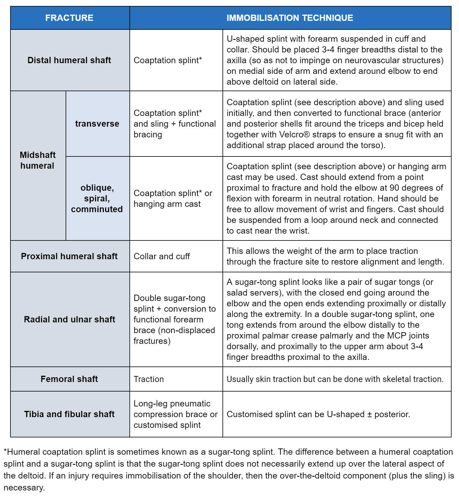
Consider venous thromboembolism prophylaxis according to current guidance.[73]
In older patients, consider the possibility of osteoporosis as an underlying cause of the fracture; investigate and manage this according to your local protocols, with referral to orthogeriatrics as necessary. See Prevention.
Suspect child maltreatment if a child has one or more fractures in the absence of a medical condition that predisposes to fragile bones and without a suitable explanation for the injury.[74] Follow your local safeguarding protocol or consult with child protection services. See Child abuse.
Practical tip
Also consider maltreatment in a child presenting with an injury that has a high association with maltreatment, such as a spiral fracture in a non-ambulatory child.
Provide appropriate analgesia: long bone fractures are typically associated with moderate to severe pain.
The type and dose of analgesia will vary with the amount of pain the patient is experiencing, the type and severity of injury, and other modifying factors (e.g., age, comorbidities, allergies). In the UK, the National Institute for Health and Care Excellence (NICE) recommends for the immediate management of pain in adults:[12]
Oral paracetamol for mild pain
Oral paracetamol and codeine for moderate pain
Intravenous paracetamol supplemented with intravenous morphine titrated to effect for severe pain.
In frail or older adults, use intravenous opioids with caution and do not offer non-steroidal anti-inflammatory drugs (NSAIDs). NSAIDs may be used as supplementary pain relief in other adults, who are not frail or elderly.[12]
In the UK, NICE recommends for the initial management of pain in children (under 16s) with suspected long bone fractures:[12]
Oral ibuprofen, or oral paracetamol, or both for mild to moderate pain
Intranasal or intravenous opioids for moderate to severe pain (use intravenous opioids if intravenous access has been established).
In practice, offer the patient with a femoral shaft fracture a femoral nerve block, and the patient with a proximal femoral fracture a fascia iliaca block.
In the UK, NICE recommends considering a femoral nerve block or fascia iliaca block in the emergency department for children (under 16s) with displaced femoral fractures.[12]
More info: Femoral nerve blocks in adults
In patients with a femoral shaft fracture who are awaiting surgical intervention, a femoral nerve block may provide superior anaesthesia to a fascia iliaca compartment block, or to isolated parenteral morphine.[75][76] However, for adult femoral shaft fractures, there is very little evidence to inform the use of this technique. One concern has been that a femoral nerve block might mask the symptoms of a developing compartment syndrome.
One randomised trial compared intravenous fentanyl with femoral nerve block prior to spinal anaesthesia for surgical intervention for femoral shaft fracture. Femoral nerve block was found to have better patient acceptance, to be associated with lower pain ratings, and to allow better positioning for spinal anaesthesia.[77] One review found no evidence to suggest that femoral nerve block delayed the diagnosis of compartment syndrome.[78]
Consider regional anaesthesia (haematoma block or peripheral nerve blockade), by healthcare professionals trained in the technique, when reducing a dorsally displaced radial fracture in adults in the emergency department.[12]
Do not give gas and air (nitrous oxide and oxygen) on its own in the emergency department for these fractures.[12]
Photograph the open fracture wound when it is first exposed for care, before debridement takes place.[45][46]
Keep the photographs in the patient’s records.
Follow your local protocol regarding taking, handling, and storing photographs and using them for clinical decision making.
In the emergency department before referral for debridement:[45][46]
Do not irrigate open fractures of long bones
Prior to formal debridement the wound should be handled only to remove gross contamination and to allow photography; ‘mini-washouts’ outside the operating theatre environment are not indicated[45]
Consider using a saline-soaked dressing covered with an occlusive layer
Administer prophylactic intravenous antibiotics, ideally within 1 hour of injury.[72]
Provide tetanus toxoid immunisation, if needed (see below).[79]
More info: Tetanus toxoid immunisation
Consider providing a tetanus toxoid immunisation. This may involve a booster dose in a patient who has received an adequate priming course, but whose last dose was more than 5-10 years ago. A patient who has not received an adequate priming course or is of uncertain immunisation status, or if there is heavy wound contamination, should receive intramuscular tetanus immunoglobulin and a reinforcing dose of vaccine.[79]
Realign and splint the limb.[45]
Consider referral for debridement, fixation, and cover of an open fracture by consultants in orthopaedic and plastic surgery.[46] Debridement should be performed:[46]
Immediately for highly contaminated open fractures
Within 12 hours of the injury for high energy open fractures that are not highly contaminated
Within 24 hours of the injury for all other open fractures.
Fixation and definitive soft-tissue cover can be performed at the same time as debridement, if the timings recommended above allow, or otherwise within 72 hours of the injury.[46] Use a temporary dressing that avoids wound desiccation and minimises the number of dressing changes after wound excision if immediate definitive soft-tissue cover has not been performed.[46]
Patients with open fractures should be treated in a setting that can provide orthoplastic care.[45]
Open fractures are often obvious, but sometimes an apparently minor surface wound belies severe injury below or nearby (bone can piston leading to a wound near the site of injury). Therefore, any fracture associated with an overlying or nearby soft-tissue injury, even an apparently innocuous minor wound, needs to be treated as an open fracture until shown otherwise.
A decision may need to be made about whether to perform limb salvage or amputation. This will require multidisciplinary assessment of the patient involving orthopaedic and plastic surgeons, a rehabilitation specialist, the patient, and their family or carers.[46]
Haemorrhage
Control frank haemorrhage with direct pressure or a tourniquet. Do not use blind clamping of bleeding.[47] Your local protocol should include combined review and decision-making in person by consultant surgeons skilled in vascular repair and skeletal trauma. The ischaemic limb should be revascularised within four hours from injury.[47]
For patients with severe acute haemorrhage, consider antifibrinolytics (e.g., tranexamic acid). These agents have been shown to increase survival.[80][81] Delay in administration reduces their benefit; in a meta-analysis of data from patients with traumatic bleeding or post-partum haemorrhage, delays in administration of tranexamic acid were associated with reduced survival (survival benefit decreasing by about 10% for every 15 minutes of treatment delay until 3 hours, after which there was no benefit).[82]
Distal humeral shaft fractures
Treat a patient who is stable and has an isolated, non-displaced humeral shaft fracture with splint immobilisation, ice, and analgesia (see Analgesia section, above).
Consult with an orthopaedic surgeon.
Most displaced humeral shaft fractures heal well with non-operative management (i.e., coaptation [or sugar-tong] splinting). Surgery is required if fracture alignment is unacceptable after closed reduction.
Midshaft humeral fracture
Splint the fracture and refer the patient to an orthopaedic surgeon for definitive treatment. Closed midshaft humeral fractures tend to heal fairly well with non-operative management. Treat a transverse fracture initially with a coaptation (or sugar-tong) splint and sling, and subsequently with functional bracing. A hanging arm cast or coaptation splint may be required for oblique, spiral, or comminuted fractures, which typically require traction.
Arrange physiotherapy with early mobilisation to restore function and minimise the chance of adhesive capsulitis of the shoulder. Fractures in which adequate positioning cannot be achieved/maintained, or which are grossly unstable, should be treated operatively.
Proximal humeral shaft fractures
Offer adults with an uncomplicated displaced proximal fracture of the humerus analgesia (see Analgesia section, above) and immobilisation in a collar and cuff. Refer for physiotherapy and arrange a non-urgent referral to orthopaedics.
Consider surgery if there is an open wound, tenting of the skin, vascular injury, fracture dislocation, rotator cuff tear, or a split of the humeral head.[12][83] Refer urgently to orthopaedics in this case.
Arrange emergency orthopaedic and vascular surgery consultations for any patient with suspected neurovascular injury.
More info: Surgery versus conservative management of proximal humerus fracture
One Cochrane review of 10 trials (717 participants) concluded there is high- or moderate-certainty evidence that, compared with non-surgical treatment, surgery does not result in a better outcome at 1 and 2 years after injury for people aged 60 and over with displaced proximal humeral fractures. A surgical approach may increase the need for subsequent surgery.[83] There is insufficient evidence from randomised controlled trials to compare surgical versus non-surgical approaches for people aged under 60 years or those with high-energy trauma, two-part tuberosity fractures, or less common fractures such as fracture dislocations or articular surface fractures.[83] Close collaboration with an experienced orthopaedic surgeon is recommended.
Initial treatment of radial fractures includes placement of a splint and urgent orthopaedic referral.
Fracture involving the proximal third of the ulna plus associated dislocation of the radial head (Monteggia fracture) requires urgent orthopaedic consultation for open reduction internal fixation (ORIF) surgery. Long-term complications include heterotopic ossification at the elbow.[84]
A sugar-tong splint is recommended for initial immobilisation of most forearm fractures; however, a double sugar-tong splint would be used in Monteggia fractures (or other elbow fractures).
[Figure caption and citation for the preceding image starts]: Sugar-tong splintPhilip Cohen [Citation ends]. [Figure caption and citation for the preceding image starts]: Double sugar-tong splintPhilip Cohen [Citation ends].
Distal radius fractures - adults
In patients with a stable fracture of the distal radius, consider early mobilisation from a removable support if pain allows.[56]
Consider regional anaesthesia (haematoma block, or peripheral nerve blockade), by healthcare professionals trained in the technique, when reducing dorsally displaced distal radius fractures (Colles fracture) in adults.[12][56] Do not use gas and air (nitrous oxide and oxygen) on its own for this purpose.[12]
Consider manipulation and a plaster cast in adults with dorsally displaced distal radius fractures.[12]
When using a plaster cast, the wrist should be in neutral flexion with 3-point moulding used to hold the fracture and not forced palmar flexion.[56]
Consider removing the cast and starting mobilisation 4 weeks after injury.[56]
Surgical fixation is sometimes needed for dorsally displaced distal radius fractures. When surgery is required for a distal radius fracture, the National Institute for Health and Care Excellence (NICE) and the British Orthopaedic Association recommend that it should be performed:[12][56]
Within 72 hours of the injury for an intra-articular fracture
Within 7 days of the injury for an extra-articular fracture.
In patients aged 65 years or older, consider non-operative treatment as the primary treatment for dorsally displaced distal radius fractures, unless there is significant deformity or neurological compromise.[56] Consider whether patients under 65 years will benefit from surgical reconstruction.
Surgical fixation may involve K-wire fixation or open reduction and internal fixation if this is not possible.[12][56] Offer K-wire fixation if no fracture of the articular surface of the radial carpal joint is detected, or if displacement of the radial carpal joint can be reduced by closed manipulation.[12]
When surgery is required for a re-displacement of distal radius fracture, NICE and the British Orthopaedic Association recommend that it should be performed within 72 hours of the decision to operate.[12][56]
Assess the patient for falls risk and bone health and refer as appropriate for any follow-up needed.[56] Explain to the patient what to expert about recovery and returning to normal activities, such as work, education, or driving.[56]
Distal radius fractures - children
Do not use a rigid cast for torus fractures of the distal radius.[12] A soft cast or bandaging may be used instead.[85] Discharge children with a torus fracture after initial assessment; further review is usually not needed.[12]
In children, early, definitive manipulation and casting without admission is the standard of care:[57]
Manipulation of a child’s forearm fracture should be performed by competent orthopaedic practitioners.
Manipulation of a child’s forearm fracture should be followed by orthogonal x-rays.
Assess the neurovascular status of the limb prior to discharge.
Provide oral analgesia to take home, along with information leaflets including information on any red flag symptoms, such as the cast being too tight (causing pain and swelling, which could create a compartment syndrome), or nerve symptoms such as pins and needles or loss of motor function.
A documented review of the case and images by a consultant orthopaedic surgeon should occur within 48 hours of injury.
For a child with a dorsally displaced distal radius fracture, who has undergone manipulation, consider a below-elbow plaster cast or K-wire fixation if the fracture is completely displaced.[12]
Explain to the patient what to expert about recovery and returning to normal activities such as education.[56]
Arrange immediate orthopaedic consultation while carrying out a thorough trauma evaluation and instituting advanced trauma or cardiovascular life support pathways.
In the pre-hospital setting, consider a traction splint or strap the limb to the adjacent leg to provide a splint for a suspected femoral fracture.[12][46]
Intramedullary nailing is the preferred treatment for most femoral shaft fractures.
Aim for immediate unrestricted weight-bearing as tolerated after surgery.[12]
Admit a child with a femoral shaft fracture .[12]
In the UK, NICE recommends that treatment should be based on the child’s age and weight. See table below.[12] In practice, consider patient factors and decide which treatment option is most suitable.
Child’s age/weight | Treatment |
|---|---|
Prematurity and birth injuries | Simple padded splint |
0 to 6 months | Pavlik harness or gallows traction |
3 to 18 months (but not in children over 15 kg) | Gallows traction |
1 to 6 years | Straight leg skin traction (becomes impractical in children over 25 kg) with possible conversion to hip spica cast to enable early discharge |
4 to 12 years (but not in children over 50 kg) | Elastic intramedullary nail |
11 years to skeletal maturity (weight more than 50 kg) | Elastic intramedullary nails supplemented by end-caps, lateral-entry antegrade rigid intramedullary nail, or submuscular plating |
In children with femoral fractures, address any concerns about non-accidental injury before discharge (particularly in children who are not yet walking or talking).[12]
Suspect child maltreatment if a child has one or more fractures in the absence of a medical condition that predisposes to fragile bones and without a suitable explanation for the injury.[74] Follow your local safeguarding protocol or consult with child protection services. See Child abuse.
Arrange immediate orthopaedic consultation for a patient with a displaced, comminuted, and open fracture.
Immobilise the fracture with a splint and provide adequate analgesia (see Analgesia section, above).
The treatment for shaft fractures is intramedullary nailing. More proximal and more distal fractures may require open reduction internal fixation (ORIF) surgery.
A closed fracture that is non-displaced and not comminuted can initially be treated with non-weight bearing and splint immobilisation, with subsequent conversion to a long leg cast, although functional bracing for truly non-displaced tibial shaft fractures is commonly used.[86][87]
Distal tibial fractures can be difficult to manage due to limited soft-tissue coverage, poor vascularity of the area, and proximity of the fracture to the ankle joint.[88] Treatment options include intramedullary nail fixation, plate-and-screw fixation, and external fixation; however, there is a lack of consensus on the best option. In a multicentre trial using a disability rating at 6 and 12 months as a measure, there was found to be no significant difference between intramedullary nail fixation and locking plate fixation.[88]
An isolated fibular fracture usually heals well with conservative care (initial non-weight bearing, followed by transition to long leg walking cast, cast boot, or compression brace). [Figure caption and citation for the preceding image starts]: Posterior leg splintPhilip Cohen [Citation ends].
Stress fractures of the upper limb
Stress fractures of the upper limb are generally treated with relative rest, analgesia (see Analgesia section, above), and a physical rehabilitation programme.
Stress fractures of the lower limb
Femoral stress fractures generally heal well with pain-free non-impact cross-training and addressing of underlying risk factors.
A patient suspected of having a femoral neck stress fracture should be made non-weight bearing immediately. Refer the patient for urgent x-rays of the hip and proximal femur.
If the films reveal a tension side fracture, a frank fracture line, or a displaced fracture, arrange an urgent orthopaedic referral for consideration of operative intervention.
If the films reveal sclerosis at the compression side, consider following up the patient with serial x-rays and having them progress to partial then full weight bearing as tolerated. Seek advice from an orthopedic consultant.
If the films are negative (common early on in the evolution of the fracture), but an MRI is positive, conservative management is reasonable. Full return to impact activity can take several months.[66]
Treat posteromedial tibial stress fractures with modified weight bearing as tolerated and cessation of impact activity. Pain-free non-impact cross-training (deep-water pool running, exercise biking, etc.) can be used to maintain fitness. Some studies have shown that the use of a pneumatic compression brace may allow the fracture to heal faster so that the patient can return to impact activity sooner.[89][90] Addressing biomechanical issues (e.g., over-pronation), ensuring proper footwear, and preventing over-training are important to prevent recurrences.
Stress fractures of the fibula are uncommon but typically occur in runners and ballet dancers. Initial x-ray findings may be negative, but MRI (or triple phase bone scan) can demonstrate the fracture earlier. Treatment includes modified activity and transition to weight bearing as tolerated. As with other stress fractures, addressing training errors and other potentially modifiable risk factors is important, as is assessing for the possibility of eating disorders and related conditions.[91]
Use of this content is subject to our disclaimer