Images and videos
Images


Long bone fracture
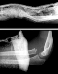
Radiographs showing dislocated radial head with distal third bone forearm fractures
Peter VK, Emerg Med J 2002;19:88-9; used with permission
See this image in context in the following section/s:

Long bone fracture
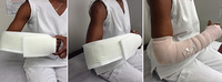
Sugar-tong splint
Philip Cohen
See this image in context in the following section/s:

Long bone fracture
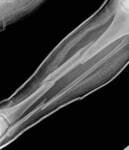
X-ray showing a segmental fracture of the tibia and fibula
See this image in context in the following section/s:
Long bone fracture
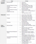
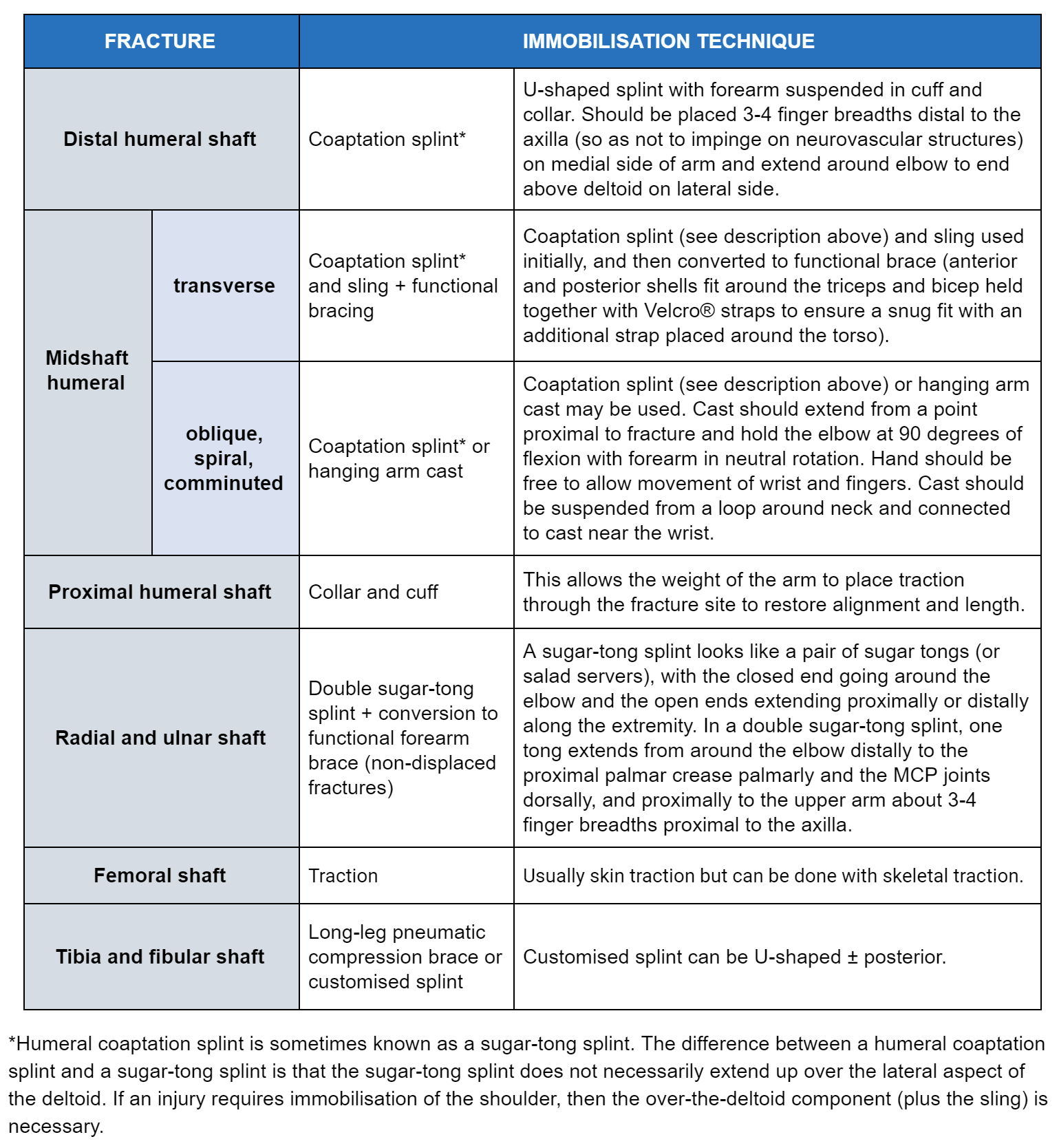
Recommended immobilisation techniques for long bone fractures
Created by BMJ Knowledge Centre
See this image in context in the following section/s:

Long bone fracture
Bilateral insufficiency lesions in proximal femora in a 63-year-old woman taking weekly alendronate
See this image in context in the following section/s:

Long bone fracture
BMJ Rapid Recommendations: low-intensity pulsed ultrasound (LIPUS) for bone healing
See this image in context in the following section/s:

Long bone fracture
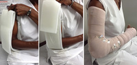
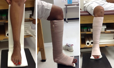
Double sugar-tong splint
Philip Cohen
See this image in context in the following section/s:
Long bone fracture
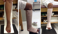
Posterior leg splint
Philip Cohen
See this image in context in the following section/s:
Long bone fracture
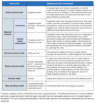
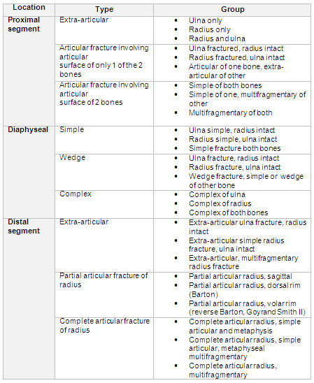
OTA classification of radius and ulna fractures - locations, types, and groups
See this image in context in the following section/s:
Use of this content is subject to our disclaimer