Images and videos
Images


Open-angle glaucoma
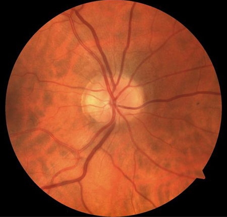
Fundus photograph of normal optic nerve head
Collection of Robert B. Avery, MD, PhD
See this image in context in the following section/s:
Open-angle glaucoma
The progressive journey from normal vision to blindness in glaucoma. There is a transition over time from normal visual function to blindness in patients with glaucoma. (A) Clinical examination or fundus photography can demonstrate and document the progression of optic disc cupping and neuroretinal rim thinning over time, that develops secondary to retinal ganglion cell loss. (B) Optical coherence tomography imaging quantifies changes in the thickness of the innermost layer of the retina around the optic disc and macula region, which comprise retinal ganglion cells and their axons, and can compare these measurements to normative databases. This enables detection and monitoring of structural changes at the optic nerve head and macula that might have developed due to glaucomatous injury. Structural changes often precede deficits in visual function and therefore optical coherence tomography imaging facilitates the detection of glaucoma at an early stage of disease. (C) Visual field testing allows for the detection and monitoring of impairment of visual function during the disease course. Early glaucoma is often asymptomatic as there is a threshold of retinal ganglion cell loss below which functional damage might not be detectable. (D) Even in the presence of substantial visual field defects, patients with glaucoma might remain asymptomatic as the brain can fill in the perceived picture using saccades and sensory inputs from the fellow eye. This means that patients might feel that their vision is normal until the very late stages of disease
Jayaram H, et al. Lancet 2023 Nov 11; 402(10414): 1788-801. doi: 10.1016/S0140-6736(23)01289-8. Epub 2023 Sep 21; used with permission
See this image in context in the following section/s:
Open-angle glaucoma
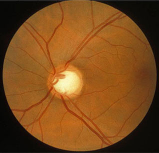
Photograph showing optic disc cupping. An increase in cup-to-disc ratio over time may indicate glaucoma.
Collection of Robert B. Avery, MD, PhD
See this image in context in the following section/s:
Use of this content is subject to our disclaimer