Despite EM being a frequently mild and self-limiting disease, a thorough history and clinical examination are essential to determine if there are any triggers that can be avoided in the future.[41]Soares A, Sokumbi O. Recent updates in the treatment of erythema multiforme. Medicina (Kaunas). 2021 Sep 1;57(9):921.
https://www.ncbi.nlm.nih.gov/pmc/articles/PMC8467974
http://www.ncbi.nlm.nih.gov/pubmed/34577844?tool=bestpractice.com
More serious mucocutaneous skin diseases such as Stevens-Johnson syndrome (SJS) and toxic epidermal necrolysis (TEN) also need to be ruled out.[3]Grünwald P, Mockenhaupt M, Panzer R, et al. Erythema multiforme, Stevens-Johnson syndrome/toxic epidermal necrolysis - diagnosis and treatment. J Dtsch Dermatol Ges. 2020 Jun;18(6):547-53.
http://www.ncbi.nlm.nih.gov/pubmed/32469468?tool=bestpractice.com
EM minor has cutaneous manifestations only, while EM major has cutaneous manifestations and involves 1 or more mucosal sites.
The lesions of both EM minor and EM major involve <10% total body surface area.
History
A detailed history of recent infection, prior recurrences, and new drug administration is necessary. Causes should be carefully investigated before any patient is labelled as having an idiopathic cause.[2]Sokumbi O, Wetter DA. Clinical features, diagnosis, and treatment of erythema multiforme: a review for the practicing dermatologist. Int J Dermatol. 2012 Aug;51(8):889-902.
http://onlinelibrary.wiley.com/doi/10.1111/j.1365-4632.2011.05348.x/full
http://www.ncbi.nlm.nih.gov/pubmed/22788803?tool=bestpractice.com
[41]Soares A, Sokumbi O. Recent updates in the treatment of erythema multiforme. Medicina (Kaunas). 2021 Sep 1;57(9):921.
https://www.ncbi.nlm.nih.gov/pmc/articles/PMC8467974
http://www.ncbi.nlm.nih.gov/pubmed/34577844?tool=bestpractice.com
Infections: the most commonly associated infections are herpes simplex virus (HSV) infection and infections caused by Mycoplasma pneumoniae.[39]Amode R, Ingen-Housz-Oro S, Ortonne N, et al. Clinical and histologic features of mycoplasma pneumoniae-related erythema multiforme: a single-center series of 33 cases compared with 100 cases induced by other causes. J Am Acad Dermatol. 2018 Jul;79(1):110-7.
http://www.ncbi.nlm.nih.gov/pubmed/29559400?tool=bestpractice.com
Other infectious agents reported to trigger EM include cytomegalovirus, Epstein-Barr virus, SARS-CoV-2, hepatitis B virus, hepatitis C virus, influenza virus, HIV, herpes zoster, gardnerella, histoplasmosis (with concomitant erythema nodosum), coccidioidomycosis, orf (a disease of sheep and goats caused by a parapox virus that can be transmitted to humans), and syphilis.[2]Sokumbi O, Wetter DA. Clinical features, diagnosis, and treatment of erythema multiforme: a review for the practicing dermatologist. Int J Dermatol. 2012 Aug;51(8):889-902.
http://onlinelibrary.wiley.com/doi/10.1111/j.1365-4632.2011.05348.x/full
http://www.ncbi.nlm.nih.gov/pubmed/22788803?tool=bestpractice.com
[7]Ma JH, Smith S, Gordon LA. Acute HIV infection presenting as erythema multiforme in a 45-year-old heterosexual man. Med J Aust. 2015 Mar 16;202(5):273-5.
https://www.mja.com.au/journal/2015/202/5/acute-hiv-infection-presenting-erythema-multiforme-45-year-old-heterosexual-man
[10]Rossi L, Tiecco G, Venturini M, et al. Human orf with immune-mediated reactions: a systematic review. Microorganisms. 2023 Apr 27;11(5):1138.
https://www.ncbi.nlm.nih.gov/pmc/articles/PMC10224112
http://www.ncbi.nlm.nih.gov/pubmed/37317112?tool=bestpractice.com
[11]Kishore BN, Ankadavar NS, Kamath GH, et al. Varicella zoster with erythema multiforme in a young girl: a rare association. Indian J Dermatol. 2014 May;59(3):299-301.
http://www.e-ijd.org/article.asp?issn=0019-5154;year=2014;volume=59;issue=3;spage=299;epage=301;aulast=Kishore
http://www.ncbi.nlm.nih.gov/pubmed/24891667?tool=bestpractice.com
[12]Kasuya A, Sakabe J, Kageyama R, et al. Successful differentiation of herpes zoster-associated erythema multiforme from generalized extension of herpes by rapid polymerase chain reaction analysis. J Dermatol. 2014 Jun;41(6):542-4.
http://www.ncbi.nlm.nih.gov/pubmed/24909215?tool=bestpractice.com
[13]Olut AI, Erkek E, Ozunlu H, et al. Erythema multiforme associated with acute hepatitis B virus infection. Clin Exp Dermatol. 2006 Jan;31(1):137-8.[14]Saleh W, Alharbi H, Cha S. Increased prevalence of erythema multiforme in patients with COVID-19 infection or vaccination. Sci Rep. 2024 Feb 2;14(1):2801.
https://www.ncbi.nlm.nih.gov/pmc/articles/PMC10837137
http://www.ncbi.nlm.nih.gov/pubmed/38307870?tool=bestpractice.com
[15]Joseph RH, Haddad FA, Matthews AL, et al. Erythema multiforme after orf virus infection: a report of two cases and literature review. Epidemiol Infect. 2015 Jan;143(2):385-90.
https://www.cambridge.org/core/journals/epidemiology-and-infection/article/div-classtitleerythema-multiforme-after-orf-virus-infection-a-report-of-two-cases-and-literature-reviewdiv/324F9FDF41A60A2218CE2AEE7F78B25D/core-reader
http://www.ncbi.nlm.nih.gov/pubmed/24810660?tool=bestpractice.com
[16]Chiang MC, Chiang FC, Chang YT, et al. Erythema multiforme caused by treponema pallidum in a young patient with human immunodeficiency virus infection. J Clin Microbiol. 2010 Jul;48(7):2640-2.
https://jcm.asm.org/content/48/7/2640.long
http://www.ncbi.nlm.nih.gov/pubmed/20504989?tool=bestpractice.com
[37]Bitterman R, Oren I, Geffen Y, et al. Prolonged fever and splinter hemorrhages in an immunocompetent traveler with disseminated histoplasmosis. J Travel Med. 2013 Jan-Feb;20(1):57-9.
https://academic.oup.com/jtm/article/20/1/57/1817330
http://www.ncbi.nlm.nih.gov/pubmed/23279234?tool=bestpractice.com
[38]Bennardo L, Nisticò SP, Dastoli S, et al. Erythema multiforme and COVID-19: what do we know? Medicina (Kaunas). 2021 Aug 16;57(8):828.
https://www.ncbi.nlm.nih.gov/pmc/articles/PMC8401222
http://www.ncbi.nlm.nih.gov/pubmed/34441034?tool=bestpractice.com
Drugs: associated medications include certain antibiotics; docetaxel or paclitaxel; immune checkpoint inhibitors; sorafenib; tumour necrosis factor (TNF)-alpha inhibitors; antimalarials; hydroxychloroquine; lenalidomide; methotrexate; anticonvulsants; statins; bisphosphonates; non-steroidal anti-inflammatory drugs (NSAIDs); metamizole; oral contraceptives; imiquimod; lidocaine; triclocarban; barium contrast.[1]Lerch M, Mainetti C, Terziroli Beretta-Piccoli B, Harr T. Current perspectives on erythema multiforme. Clin Rev Allergy Immunol. 2018 Feb;54(1):177-84.
http://www.ncbi.nlm.nih.gov/pubmed/29352387?tool=bestpractice.com
[18]Borrás-Blasco J, Navarro-Ruiz A, Borrás C, et al. Adverse cutaneous reactions induced by TNF-alpha antagonist therapy. South Med J. 2009 Nov;102(11):1133-40.
http://www.ncbi.nlm.nih.gov/pubmed/19864977?tool=bestpractice.com
[19]Lenalidomide: Stevens-Johnson syndrome. Prescrire Int. 2010 Jun;19(107):125.
http://www.ncbi.nlm.nih.gov/pubmed/20740722?tool=bestpractice.com
[20]Ballester I, Guijarro J, Silvestre JF, et al. Erythema multiforme induced by imiquimod 5% cream. Int J Dermatol. 2014 Jul;53(7):e347-8.
http://www.ncbi.nlm.nih.gov/pubmed/24602041?tool=bestpractice.com
[21]Sai Keerthana PC, Anila KN, Reshma R. Naproxen induced erythema multiforme - a rare case report. Int J Pharm and Pharmaceutical Sci. 2017;9:294-5.
https://innovareacademics.in/journals/index.php/ijpps/article/viewFile/14903/9961
[22]Rodríguez-Pazos L, Sánchez-Aguilar D, Rodríguez-Granados MT, et al. Erythema multiforme photoinduced by statins. Photodermatol Photoimmunol Photomed. 2010 Aug;26(4):216-8.
http://www.ncbi.nlm.nih.gov/pubmed/20626826?tool=bestpractice.com
[23]Rodríguez-Pazos L, Gómez-Bernal S, Rodríguez-Granados MT, et al. Photodistributed erythema multiforme. Actas Dermosifiliogr. 2013 Oct;104(8):645-53.
https://www.actasdermo.org/en-photodistributed-erythema-multiforme-articulo-S1578219013001728
http://www.ncbi.nlm.nih.gov/pubmed/23962583?tool=bestpractice.com
[24]Utsunomiya A, Oyama N, Iino S, et al. A case of erythema multiforme major developed after sequential use of two immune checkpoint inhibitors, nivolumab and ipilimumab, for advanced melanoma: possible implication of synergistic and/or complementary immunomodulatory effects. Case Rep Dermatol. 2018 Jan 18;10(1):1-6.
https://www.ncbi.nlm.nih.gov/pmc/articles/PMC5836162
http://www.ncbi.nlm.nih.gov/pubmed/29515387?tool=bestpractice.com
[25]de Arruda JA, Silva P, Amaral MB, et al. Erythema multiforme induced by alendronate sodium in a geriatric patient: a case report and review of the literature. J Clin Exp Dent. 2017 Jul;9(7):e929-33.
https://www.ncbi.nlm.nih.gov/pmc/articles/PMC5549594
http://www.ncbi.nlm.nih.gov/pubmed/28828163?tool=bestpractice.com
[26]Abou Assalie N, Durcan R, Durcan L, et al. Hydroxychloroquine-induced erythema multiforme. J Clin Rheumatol. 2017 Mar;23(2):127-8.
https://www.ncbi.nlm.nih.gov/pmc/articles/PMC5321779
[27]Mantovani A, Álvares-Da-Silva MR. Anaphylaxis preceded by erythema multiforme with sorafenib: first case report. Ann Hepatol. 2019 Sep - Oct;18(5):777-9.
https://www.sciencedirect.com/science/article/pii/S1665268119300997?via%3Dihub
http://www.ncbi.nlm.nih.gov/pubmed/31085038?tool=bestpractice.com
However, this list is not exhaustive and you should check your local drug information source. Photo-distributed lesions have been noted with phenylbutazone, triclocarban, paclitaxel, and statins.[22]Rodríguez-Pazos L, Sánchez-Aguilar D, Rodríguez-Granados MT, et al. Erythema multiforme photoinduced by statins. Photodermatol Photoimmunol Photomed. 2010 Aug;26(4):216-8.
http://www.ncbi.nlm.nih.gov/pubmed/20626826?tool=bestpractice.com
[23]Rodríguez-Pazos L, Gómez-Bernal S, Rodríguez-Granados MT, et al. Photodistributed erythema multiforme. Actas Dermosifiliogr. 2013 Oct;104(8):645-53.
https://www.actasdermo.org/en-photodistributed-erythema-multiforme-articulo-S1578219013001728
http://www.ncbi.nlm.nih.gov/pubmed/23962583?tool=bestpractice.com
Vaccines and allergens: vaccines against organisms such as hepatitis B, smallpox, varicella, meningococcus, human papillomavirus, and SARS-CoV-2 have been known to elicit EM.[14]Saleh W, Alharbi H, Cha S. Increased prevalence of erythema multiforme in patients with COVID-19 infection or vaccination. Sci Rep. 2024 Feb 2;14(1):2801.
https://www.ncbi.nlm.nih.gov/pmc/articles/PMC10837137
http://www.ncbi.nlm.nih.gov/pubmed/38307870?tool=bestpractice.com
[28]Chahal D, Aleshin M, Turegano M, et al. Vaccine-induced toxic epidermal necrolysis: a case and systematic review. Dermatol Online J. 2018 Jan 15;24(1):13030/qt7qn5268s.
https://escholarship.org/uc/item/7qn5268s
http://www.ncbi.nlm.nih.gov/pubmed/29469759?tool=bestpractice.com
[40]Yousefian M, Khadivi A. Occurrence of erythema multiforme following COVID-19 vaccination: a review. Clin Exp Vaccine Res. 2023 Apr;12(2):87-96.
https://www.ncbi.nlm.nih.gov/pmc/articles/PMC10193109
http://www.ncbi.nlm.nih.gov/pubmed/37214146?tool=bestpractice.com
Allergic response to contact allergens, such as tattoos, can also trigger EM.[29]Allione A, Dutto L, Castagna E, et al. Erythema multiforme caused by tattoo: a further case. Intern Emerg Med. 2011 Jun;6(3):263-5.
https://link.springer.com/article/10.1007%2Fs11739-010-0394-5
The lesions typically appear a few days after the trigger. Some lesions will initially clearly resemble target lesions; others will evolve from small erythematous plaques. The lesions have a rapid onset and usually increase in number over 4 to 7 days. They can cause general discomfort but are not itchy until they start to heal. Oral mucosal involvement may be particularly painful for the patient, and in more severe cases causes a reduced ability to take fluids and food.
Clinical examination
EM presents with typical target lesions (annular erythematous rings with an outer erythematous zone and central blister sandwiching a zone of normal skin tone) and atypical targetoid papules (no central blistering). The clinical pattern of the lesions is the most important diagnostic tool, with the characteristic target lesions commonly found in a symmetrical distribution on the extremities. Targetoid lesions are more common centripetally.[2]Sokumbi O, Wetter DA. Clinical features, diagnosis, and treatment of erythema multiforme: a review for the practicing dermatologist. Int J Dermatol. 2012 Aug;51(8):889-902.
http://onlinelibrary.wiley.com/doi/10.1111/j.1365-4632.2011.05348.x/full
http://www.ncbi.nlm.nih.gov/pubmed/22788803?tool=bestpractice.com
[3]Grünwald P, Mockenhaupt M, Panzer R, et al. Erythema multiforme, Stevens-Johnson syndrome/toxic epidermal necrolysis - diagnosis and treatment. J Dtsch Dermatol Ges. 2020 Jun;18(6):547-53.
http://www.ncbi.nlm.nih.gov/pubmed/32469468?tool=bestpractice.com
[4]Bastuji-Garin S, Rzany B, Stern RS, et al. Clinical classification of cases of toxic epidermal necrolysis, Stevens-Johnson syndrome, and erythema multiforme. Arch Dermatol. 1993 Jan;129(1):92-6.
http://www.ncbi.nlm.nih.gov/pubmed/8420497?tool=bestpractice.com
[5]Assier H, Bastuji-Garin S, Revuz J, et al. Erythema multiforme with mucous membrane involvement and Stevens-Johnson syndrome are clinically different disorders with distinct causes. Arch Dermatol. 1995 May;131(5):539-43.
http://www.ncbi.nlm.nih.gov/pubmed/7741539?tool=bestpractice.com
In the presence of target lesions, targetoid lesions corroborate the diagnosis of EM, when they occur rapidly. Mucosal membranes of the mouth, eyes, nose, and genitalia should also be examined for mucosal erosions seen in EM major. Some typical target lesions and minimal mucosal disease is the finding most suggestive of EM, especially in the setting of HSV infection or Mycoplasma pneumoniae infection.
A more general physical examination should also be carried out to identify any possible infectious cause. HSV infection is characterised by clustered vesicles on an erythematous base. A red tympanic membrane strongly suggests Mycoplasma pneumoniae, as do rhonchi, rales, and/or wheezes.[Figure caption and citation for the preceding image starts]: Palmar target lesionsFrom the personal collection of Nanette Silverberg, MD; used with permission [Citation ends].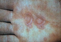 [Figure caption and citation for the preceding image starts]: Target and targetoid lesionsFrom the personal collection of Nanette Silverberg, MD; used with permission [Citation ends].
 [Figure caption and citation for the preceding image starts]: Target lesions on the face and mucosal erosions with crusting due to HSV-1 recurrenceFrom the personal collection of Nanette Silverberg, MD; used with permission [Citation ends].
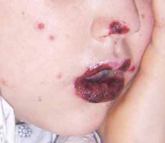
Laboratory evaluation
Most cases of EM can be diagnosed by history and clinical examination alone, and no further investigations are needed. However, if there is diagnostic uncertainty after clinical examination, a biopsy for haematoxylin and eosin staining can be performed. If the result of this biopsy is not conclusive, a biopsy for immunofluorescence can also be carried out.
If the cause of EM is not apparent from clinical examination, laboratory tests are performed in order to try to establish a cause. Since the most common infections to trigger EM are HSV and mycoplasma, the initial tests are full blood count, electrolytes, HSV serology, cold agglutinins, M pneumoniae titres, and/or chest x-ray (depending on the clinical state of the patient). If these tests are negative, then tests for other less common infectious causes are carried out. Herpes zoster-associated EM can be differentiated from generalised extension of herpes by rapid polymerase chain reaction.[12]Kasuya A, Sakabe J, Kageyama R, et al. Successful differentiation of herpes zoster-associated erythema multiforme from generalized extension of herpes by rapid polymerase chain reaction analysis. J Dermatol. 2014 Jun;41(6):542-4.
http://www.ncbi.nlm.nih.gov/pubmed/24909215?tool=bestpractice.com
HSV serology can also be useful if there have been recurrent episodes of EM but there are no specific HSV lesions found. Anti-desmoplakin antibodies have been noted in patients with recurrent EM.[36]Wetter DA, Davis MD. Recurrent erythema multiforme: clinical characteristics, etiologic associations, and treatment in a series of 48 patients at Mayo Clinic, 2000 to 2007. J Am Acad Dermatol. 2010 Jan;62(1):45-53.
http://www.jaad.org/article/S0190-9622(09)00778-6/fulltext
http://www.ncbi.nlm.nih.gov/pubmed/19665257?tool=bestpractice.com
Distinguishing Stevens-Johnson syndrome and toxic epidermal necrolysis from EM
Other more severe reaction patterns should be ruled out, including those of SJS and TEN. Severe limitation of oral intake and pain with voiding are more common with these diseases, but can be seen with EM major.[2]Sokumbi O, Wetter DA. Clinical features, diagnosis, and treatment of erythema multiforme: a review for the practicing dermatologist. Int J Dermatol. 2012 Aug;51(8):889-902.
http://onlinelibrary.wiley.com/doi/10.1111/j.1365-4632.2011.05348.x/full
http://www.ncbi.nlm.nih.gov/pubmed/22788803?tool=bestpractice.com
[3]Grünwald P, Mockenhaupt M, Panzer R, et al. Erythema multiforme, Stevens-Johnson syndrome/toxic epidermal necrolysis - diagnosis and treatment. J Dtsch Dermatol Ges. 2020 Jun;18(6):547-53.
http://www.ncbi.nlm.nih.gov/pubmed/32469468?tool=bestpractice.com
[4]Bastuji-Garin S, Rzany B, Stern RS, et al. Clinical classification of cases of toxic epidermal necrolysis, Stevens-Johnson syndrome, and erythema multiforme. Arch Dermatol. 1993 Jan;129(1):92-6.
http://www.ncbi.nlm.nih.gov/pubmed/8420497?tool=bestpractice.com
[5]Assier H, Bastuji-Garin S, Revuz J, et al. Erythema multiforme with mucous membrane involvement and Stevens-Johnson syndrome are clinically different disorders with distinct causes. Arch Dermatol. 1995 May;131(5):539-43.
http://www.ncbi.nlm.nih.gov/pubmed/7741539?tool=bestpractice.com
[9]Auquier-Dunant A, Mockenhaupt M, Maldi L, et al. Correlations between clinical patterns and causes of erythema multiforme majus, Stevens-Johnson syndrome, and toxic epidermal necrolysis: results of an international prospective study. Arch Dermatol. 2002 Aug;138(8):1019-24.
https://jamanetwork.com/journals/jamadermatology/fullarticle/478935
http://www.ncbi.nlm.nih.gov/pubmed/12164739?tool=bestpractice.com
[42]Cote B, Wechsler J, Bastuji-Garin S. Clinicopathologic correlation in erythema multiforme and Stevens-Johnson syndrome. Arch Dermatol. 1995 Nov;131(11):1268-72.
http://www.ncbi.nlm.nih.gov/pubmed/7503570?tool=bestpractice.com
SJS affects <10% total body surface area and tends to have extensive oral and genital mucosal involvement. Often, an offending drug exposure is identified. TEN demonstrates extensive denudation of the skin, generally more than 30%. If it is difficult to differentiate a suspected case of EM from SJS or TEN, 2 tests can be performed:
The Asboe-Hansen sign of TEN is typified by the physical enlargement of blisters with direct pressure applied to the top of the blister, demonstrating basal keratinocyte necrosis. SJS and TEN also have the Nikolsky's sign of skin slough on touch, not present in EM.[2]Sokumbi O, Wetter DA. Clinical features, diagnosis, and treatment of erythema multiforme: a review for the practicing dermatologist. Int J Dermatol. 2012 Aug;51(8):889-902.
http://onlinelibrary.wiley.com/doi/10.1111/j.1365-4632.2011.05348.x/full
http://www.ncbi.nlm.nih.gov/pubmed/22788803?tool=bestpractice.com
Biopsy and fresh frozen tissue assessment can demonstrate necrotic keratinocytes in SJS and TEN. Monocytic infiltrates and red blood cells are more typical on histopathology of EM; there is no epidermal necrosis and the inflammatory infiltrate is within the dermis.[2]Sokumbi O, Wetter DA. Clinical features, diagnosis, and treatment of erythema multiforme: a review for the practicing dermatologist. Int J Dermatol. 2012 Aug;51(8):889-902.
http://onlinelibrary.wiley.com/doi/10.1111/j.1365-4632.2011.05348.x/full
http://www.ncbi.nlm.nih.gov/pubmed/22788803?tool=bestpractice.com
[3]Grünwald P, Mockenhaupt M, Panzer R, et al. Erythema multiforme, Stevens-Johnson syndrome/toxic epidermal necrolysis - diagnosis and treatment. J Dtsch Dermatol Ges. 2020 Jun;18(6):547-53.
http://www.ncbi.nlm.nih.gov/pubmed/32469468?tool=bestpractice.com
[4]Bastuji-Garin S, Rzany B, Stern RS, et al. Clinical classification of cases of toxic epidermal necrolysis, Stevens-Johnson syndrome, and erythema multiforme. Arch Dermatol. 1993 Jan;129(1):92-6.
http://www.ncbi.nlm.nih.gov/pubmed/8420497?tool=bestpractice.com
[5]Assier H, Bastuji-Garin S, Revuz J, et al. Erythema multiforme with mucous membrane involvement and Stevens-Johnson syndrome are clinically different disorders with distinct causes. Arch Dermatol. 1995 May;131(5):539-43.
http://www.ncbi.nlm.nih.gov/pubmed/7741539?tool=bestpractice.com
[32]Sundram U. A review of important skin disorders occurring in the posttransplantation patient. Adv Anat Pathol. 2014 Sep;21(5):321-9.
http://www.ncbi.nlm.nih.gov/pubmed/25105934?tool=bestpractice.com
[42]Cote B, Wechsler J, Bastuji-Garin S. Clinicopathologic correlation in erythema multiforme and Stevens-Johnson syndrome. Arch Dermatol. 1995 Nov;131(11):1268-72.
http://www.ncbi.nlm.nih.gov/pubmed/7503570?tool=bestpractice.com
See Stevens-Johnson syndrome and toxic epidermal necrolysis (Diagnosis Approach).