Images and videos
Images


Assessment of chest pain
Distribution of final diagnoses in people over 35 years admitted to hospital from one US hospital emergency department with chief complaint of non-traumatic chest pain, over a 5-year period (PE, pulmonary embolism)
Created by BMJ; based on data from Kohn MA, Kwan E, Gupta M, et al. Prevalence of acute myocardial infarction and other serious diagnoses in patients presenting to an urban emergency department with chest pain. J Emerg Med. 2005 Nov;29(4):383-90
See this image in context in the following section/s:
Assessment of chest pain
Composition of the HEART score for chest pain patients in the emergency department
Six AJ, et al. Neth Heart J. 2008;16:191-6; used with permission
See this image in context in the following section/s:

Assessment of chest pain
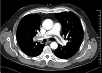
Spiral computed tomography pulmonary angiogram showing a large filling defect within the pulmonary vasculature compatible with a saddle embolus
From the collection of Professor James Brown; used with permission
See this image in context in the following section/s:

Assessment of chest pain
Varicella zoster virus
Courtesy of Daniel Eisen, MD; used with permission
See this image in context in the following section/s:
Assessment of chest pain
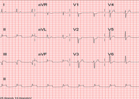
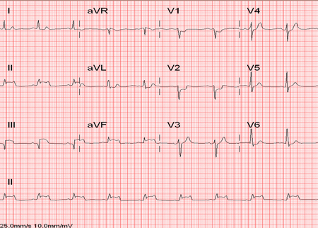
ECG showing changes of an acute inferior myocardial infarction with ST elevation in leads II, III, and aVF
From the collection of Professor James Brown; used with permission
See this image in context in the following section/s:
Assessment of chest pain
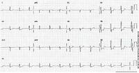
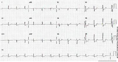
ECG showing ST depression
From the personal collection of Dr Syed W. Yusuf and Dr Iyad N. Daher, Department of Cardiology, University of Texas, Houston, TX; used with permission
See this image in context in the following section/s:
Assessment of chest pain
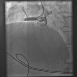
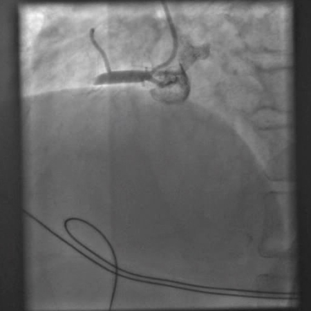
Angiogram showing occluded right artery
From the personal collection of Dr Mahi Ashwath; used with permission
See this image in context in the following section/s:
Assessment of chest pain
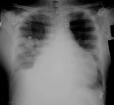
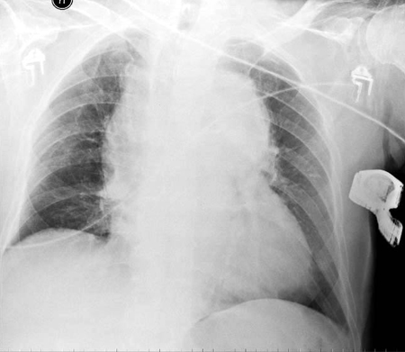
Chest x-ray showing a widened mediastinum
From the collection of Professor James Brown; used with permission
See this image in context in the following section/s:

Assessment of chest pain
Chest x-ray showing a large globular heart in a patient with pericardial tamponade
From the collection of Professor James Brown; used with permission
See this image in context in the following section/s:
Videos
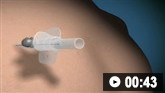 Needle decompression of tension pneumothorax animated demonstration
How to decompress a tension pneumothorax. Demonstrates insertion of a large-bore intravenous cannula into the fourth intercostal space in an adult.
 Venepuncture and phlebotomy animated demonstration
How to take a venous blood sample from the antecubital fossa using a vacuum needle.
 How to perform an ECG animated demonstration
How to record an ECG. Demonstrates placement of chest and limb electrodes.
Use of this content is subject to our disclaimer