Investigations
1st investigations to order
spine x-ray
Test
Spine radiography is the cornerstone of the diagnosis of DISH, although DISH is often an incidental finding during routine imaging.[1]
Request anteroposterior and lateral radiographs of the thoracic, lumbar, and cervical spine if DISH is suspected.[3]
The osteophytes of DISH project from the vertebral bodies as opposed to syndesmophytes, which project from the annulus fibrosus; a distinction that is useful when it occurs, but is not always easy to determine.
The osteophytes of DISH are most likely to be seen on the right anterior thoracic spine, directly across from the pulsating aorta.[1]
Lower thoracic and thoracolumbar spine involvement is common.[1]
Look for coarse, thick osteophytes that develop mostly on the right side and horizontally along the anterior longitudinal ligament.[4] These findings are often described as having a 'flowing candle wax' appearance. 'Flowing' osteophytes, at a minimum of 3-4 contiguous vertebrae, are a distinguishing feature of DISH.[34][38][39][40]
There is a lack of universally accepted validated criteria. In practice, 4 continuous flowing osteophytes is generally considered a definitive diagnosis of DISH and 3 is considered probable DISH.
Other characteristic diagnostic features include preservation of disc height and lack of significant degenerative changes at the involved vertebral segments, as well as absence of ankylosis at the facet-joint interface and absence of sacroiliac joint erosion, sclerosis, or fusion.[34]
DISH frequently co-exists with age-related disc or facet joint degenerative changes, which may be noted on radiographs.[41]
[Figure caption and citation for the preceding image starts]: X-ray images of the thoracic spine of a patient with DISH. (A–C) Posterior–anterior and (D) lateral: large right-sided flowing bridges (white arrows). Note the space between the ligament and the vertebral body (*). Thick flowing ossification of the anterior lateral ligament is shown (black arrow)Mader R, et al. RMD Open 2020; 6: e001151. doi: 10.1136; used with permission [Citation ends].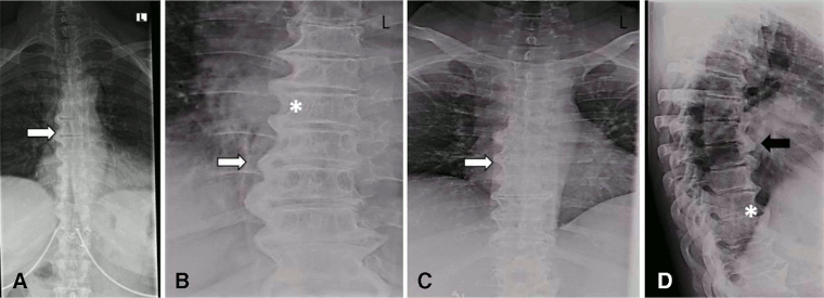
Result
may reveal presence of coarse, thick osteophytes often on the right side and horizontally along the anterior longitudinal ligament, with a 'flowing candle wax' appearance; may reveal preservation of disc height and lack of significant degenerative changes at the involved vertebral segments, absence of ankylosis at the facet-joint interface, and absence of sacroiliac joint erosion, sclerosis, or fusion
Investigations to consider
CT spine
Test
Although spine radiography is the investigation of choice for identifying characteristic bone formation, CT may also be considered.[4]
CT may be useful for detection of early changes secondary to DISH, and is a more sensitive imaging modality than x-ray in showing structural changes; however, this must be weighed against the harm of additional radiation exposure.
Criteria have been proposed to identify patients with early DISH on CT.[38] A score from 0 to 3 is assigned for each vertebral segment adjacent to a complete bone bridge, depending on presence of osteophytes, near complete bridging (<2 mm distance between bony structures), and complete bridging (full connection between two adjacent bones with abundant new bone formation).[38] The presence of <3 adjacent segments with a complete bone bridge is identified as early DISH, while the presence of ≥3 is labelled as definite DISH.[38]
[Figure caption and citation for the preceding image starts]: CT images of the thoracic spine in DISH. (A–C) Sagittal: CT scan images of anterior flowing osteophytes (arrows). (D) Coronal: dish of the thoracic spine (arrow) reconstructed from the chest CT scan. L = leftMader R, et al. RMD Open 2020; 6: e001151. doi: 10.1136; used with permission [Citation ends].
Other indications for CT include fracture detection and assessment of dysphagia.[1][3]
Request a whole-spine CT scan for patients with suspected spinal fractures. It can be challenging to identify spinal fractures in DISH on plain radiographs, due to the presence of degenerative changes and occult fracture lines, and whole-spine CT can provide a clearer view in patients with suspected spinal fractures.
A CT myelogram may be considered for cases with neurological involvement or fractures.[47] However, the evidence for this is not robust.
Result
may reveal presence of large bridging osteophytes and ossification of the anterior lateral ligament; may reveal spinal fracture
peripheral joint x-ray
Test
Order in patients with peripheral symptoms.
Result
may reveal joint space narrowing, enthesopathy, osteophytes, ossification of the tendon
MRI spine
Test
Usually non-contrast. Consider contrast-enhanced study when evaluation of the spinal column is important.
MRI can be useful to detect occult fractures, particularly in patients with neurological deficits.[1]
Helpful when evaluating for spinal fracture where sequences may show inflammatory changes secondary to a fracture.
Result
may reveal osteophytes, ossification of anterior and posterior longitudinal ligament, enthesitis of interspinous ligaments; occult fractures
ultrasound
Test
Musculoskeletal ultrasound can be used for evaluation of entheseal changes in peripheral joints.[42]
Result
may reveal entheseal changes
dual-energy x-ray absorptiometry (DXA)/whole-spine vertebral fracture assessment (VFA)
Test
Consider DXA/VFA to measure bone mineral density in patients with fracture secondary to DISH.
Note that ossification of ligaments and formation of bony bridges in patients with DISH can be mistaken for increased bone density by the DXA scan.
Result
may reveal reduced bone mineral density
quantitative computed tomography (QCT)
Test
Consider QCT as an alternative to DXA/VFA to measure bone mineral density in patients with fracture secondary to DISH. Ossification of ligaments and formation of bony bridges in patients with DISH can be mistaken for increased bone density with DXA imaging. QCT may be useful to help differentiate between cortical and trabecular bone.[44]
Result
may reveal reduced bone mineral density
video swallow study
Test
Order in people with dysphagia or dyspnoea.[1]
Result
may reveal large osteophytes compressing oesophagus or trachea
laryngoscopy and oesophagoscopy
Test
Order in people with dysphagia or dyspnoea.[1]
Result
may reveal large osteophytes compressing oesophagus or trachea
pulmonary function tests
Test
There may be an association between DISH and pulmonary function abnormalities. A restrictive spirometry pattern with evidence of extrathoracic obstruction has been noted in a cohort of heavy smokers with DISH.[43]
Result
may demonstrate a restrictive pattern
C-reactive protein
Test
Blood tests such as C-reactive protein, HLA-B27, rheumatoid factor, and antinuclear antibody levels may be useful to help exclude differentials; note, results are frequently normal in patients with DISH.
Result
normal
HLA-B27
Test
Blood tests such as HLA-B27, C-reactive protein, rheumatoid factor, and antinuclear antibody levels may be useful to help exclude differentials such as ankylosing spondylitis; note, results are frequently normal in patients with DISH.
Result
negative
rheumatoid factor
Test
Blood tests such as rheumatoid factor levels may be useful to help exclude differentials; note, results are frequently normal in patients with DISH.
Result
negative
antinuclear antibodies (ANA)
Test
Blood tests such as antinuclear antibody levels may be useful to help exclude differentials; note, results are frequently normal in patients with DISH.
Result
negative
Emerging tests
sclerostin
Use of this content is subject to our disclaimer