Images and videos
Images

Community-acquired pneumonia in children
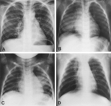
Chest radiographs confirming pneumonia. Image A: a 6-year-old girl with widespread interstitial changes in both lungs caused by S pneumoniae. Image B: a 1-year-old boy with alveolar changes in the right lower lobe caused by S pneumoniae. Image C: a 2-year-old girl with alveolar changes in the left lower lobe associated with rhinovirus. Image D: a 4-month-old girl with alveolar changes in the right upper lobe associated with parainfluenza 2 and human herpes virus
Virkki R, et al. Thorax 2002; 57: 438-41; used with permission
See this image in context in the following section/s:

Community-acquired pneumonia in children
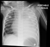
Chest x-ray of complicated pneumonia, showing opacification of the left lung field consistent with a large pleural effusion and empyema. There is associated right-sided bronchial wall thickening and consolidation
Haq IJ, et al. BMJ 2017 Mar 2; 356: j686. doi: 10.1136/bmj.j686; used with permission
See this image in context in the following section/s:
Videos
 Early inspiratory crackles
Auscultation sounds: Early inspiratory crackles
 Late inspiratory crackles (rales)
Auscultation sounds: late inspiratory crackles (rales)
Use of this content is subject to our disclaimer