History and exam
Key diagnostic factors
common
fever
Consider the possibility of pneumonia if a child presents with a fever, particularly if associated with one or more of the following: tachypnoea; chest crackles; nasal flaring; chest indrawing; cyanosis; oxygen saturation ≤95% on room air.[9][20]
One multi-centre study covering 2358 children who had radiographic evidence of pneumonia found that 91% had fever.[4] Fever >37.5°C (>99.5°F) had a likelihood ratio range of 1.7 to 1.8 for predicting radiographically confirmed CAP (sensitivity 80% to 92%, specificity 47% to 54%) in a systematic review of 23 studies involving 13,833 children with suspected pneumonia.[5]
A fever that persists for >7 days raises suspicion for empyema, and a prolonged high fever is also a typical feature of necrotising pneumonia.[2][3][9]
cough
hypoxaemia
Use pulse oximetry to check for hypoxaemia.
Hypoxaemia and signs of increased work of breathing were the two symptoms or signs most strongly correlated with radiographic evidence of pneumonia in one systematic review of 23 prospective cohort studies involving a total of 13,833 children with suspected pneumonia.[5] Oxygen saturation ≤96% on pulse oximetry was found to have a likelihood ratio of 2.8 (95% CI 2.1 to 3.6), a sensitivity of 64%, and a specificity of 77% for pneumonia. Conversely, oxygen saturation >96% was a strong predictor that the child would not have radiographic evidence of pneumonia (likelihood ratio 0.47, 95% CI 0.32 to 0.67).[5]
Arrange hospital admission for any child or infant whose oxygen saturation level indicates severe CAP. The precise threshold for hypoxaemia that warrants hospital admission varies between guidelines, so check your local protocol.
The UK National Institute of Health and Care Excellence (NICE) states that oxygen saturation <90% is a sign of severe pneumonia.[18] The British Thoracic Society paediatric CAP guideline defines oxygen saturation <92% as a sign of severe pneumonia.[9]
The national US paediatric CAP guideline recommends hospital admission for any child or infant with CAP who has sustained peripheral oxygen saturation <90% on room air, on the basis that this is predictive of failure of outpatient oral antibiotic treatment.[1]
tachypnoea
A common but non-specific sign of CAP.[1][9]
Respiratory rate (RR) >40 breaths/minute had a likelihood ratio for predicting radiographically confirmed CAP of 1.5 (95% CI 1.3 to 1.7) (sensitivity 79%, specificity 51%) in one systematic review of 23 prospective cohort studies involving a total of 13,833 children with suspected pneumonia.[5]
Be aware, however, that some children with CAP have a normal RR.[9]
A raised RR compared with age-specific norms has been found to correlate well with hypoxaemia.[21] One study found that in infants <1 year, an RR ≥70 breaths/minute had a sensitivity of 63% and specificity of 89% for hypoxaemia.[28]
Arrange hospital admission for any child with significant tachypnoea.[1] Note that tachypnoea is defined according to age-related criteria, although suggested reference ranges for different paediatric age groups vary between different sources.
The British Thoracic Society (BTS) paediatric CAP guideline defines RR (breaths/minute) of >70 in infants or >50 in older children as a sign of severe pneumonia.[9]
The UK National Institute for Health and Care Excellence (NICE) defines tachypnoea in the context of suspected pneumonia as RR (breaths/minute): >60 at age 0-5 months; >50 at age 6-12 months; >40 at ages 1-5 years.[20] The NHS England national paediatric early warning system (PEWS) uses a threshold for 5- to 12-year-olds of >25 breaths/minute for mild respiratory distress, >40 for moderate respiratory distress, and >50 for severe respiratory distress.[30] For children ≥13 years old, PEWS defines RR >25 breaths/minute as mild respiratory distress, >30 as moderate respiratory distress, and >40 as severe respiratory distress.[30]
The US national paediatric CAP guideline recommends hospital admission for any child with an RR (breaths/minute): >60 at age 0-2 months; >50 at age 2-12 months; >40 at age 1-5 years; >20 at age >5 years.[1]
increased work of breathing
Look for: suprasternal, intercostal, or subcostal retractions; nasal flaring; or head bobbing.[1][9][18]
Arrange hospital admission for any child or infant with CAP who has signs of substantially increased work of breathing (e.g., retractions, nasal flaring, use of accessory muscles).[1]
Signs of increased work of breathing and hypoxaemia were the two symptoms or signs most strongly correlated with radiographic evidence of pneumonia in one systematic review of 23 prospective cohort studies involving a total of 13,833 children with suspected pneumonia.[5] Increased work of breathing was found to have a likelihood ratio of 2.1 (95% CI 1.6 to 2.7) for predicting radiographically confirmed pneumonia.[5]
Grunting is a sign of severe disease and impending respiratory failure.[1]
abnormal auscultatory findings
Signs of CAP on auscultation may include abnormal or decreased breath sounds such as crackles, rales, crepitation, wheeze, and rhonchi.[1] One study found that crackles and bronchial breathing had a sensitivity of 75% and specificity of 57% for pneumonia.[28]
Note, however, that a systematic review of 23 studies involving a total of 13,833 children with suspected pneumonia found that no auscultatory finding was significantly associated with a radiographic diagnosis of CAP, perhaps because of the relative subjectivity of auscultatory signs and difficulty interpreting them in children.[5]
Arrange hospital admission for any child with auscultatory findings that suggest complicated CAP.[1][9] An absence of breath sounds, with a dull percussion note, is suggestive of CAP complicated by pleural effusion.[9] Fremitus is increased in uncomplicated CAP (but reduced if pleural effusion has developed).[2]
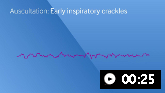
Auscultation sounds: Early inspiratory crackles
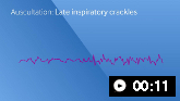
Auscultation sounds: late inspiratory crackles (rales)
uncommon
Other diagnostic factors
common
dyspnoea
tachycardia
Check the pulse rate for any signs of tachycardia.[3][9]
Tachycardia is defined according to age-related norms. It is usually considered to be 2 standard deviations above the age-standardised normal heart rate or <10th percentile for age if the child is <1 year old.
The UK National Institute for Health and Care Excellence (NICE) defines tachycardia as follows for children <5 years old:[20]
>160 bpm for infants <1 year old
>150 bpm for children aged 12-24 months
>140 bpm for children aged 2-5 years.
wheeze
Only an indicator of possible CAP if accompanied by fever and/or hypoxaemia.[1] One study of 526 children presenting to the emergency department with wheeze found that only 4.9% of those with wheeze alone had radiographic evidence of pneumonia, compared with 20.6% of those who also had both fever and hypoxaemia (oxygen saturation <92%).[24]
Wheeze on its own is a poor indicator of possible CAP and raises suspicion of an alternative diagnosis, such as viral wheeze or an exacerbation of asthma. The presence of wheeze has been found in several studies to be a negative predictor of radiographic CAP.[10][22][23]
uncommon
prolonged capillary refill time (CRT)
A CRT >2 seconds is a sign of severe disease.[9]
chest pain
abdominal pain
Is occasionally the main presenting symptom, especially in children <5 years old.[1]
vomiting
difficulty feeding
agitation or altered mental status
May be an indicator of hypoxaemia. Arrange hospital admission if present.[1]
Risk factors
strong
younger age (<2 years old)
prematurity
chronic underlying condition
Among the long-term conditions associated with a higher risk of developing CAP, and particularly complicated CAP, are: immunodeficiency; malnutrition; chronic lung disease; congenital heart disease; neurodisability; cerebral palsy; cystic fibrosis; and a history of severe and/or complicated and/or recurrent pneumonia.[1][2][3]
history of severe and/or complicated and/or recurrent pneumonia
A history of recurrent pneumonia or of severe or complicated pneumonia increases the risk of progressing to severe or complicated CAP.[3]
inhaled foreign body
An undiagnosed and retained inhaled foreign body may result in CAP or complicated CAP (e.g., atelectasis, bronchiectasis, lung abscess).[2]
indoor air pollution
overcrowded housing
Data suggest that household crowding puts young children at increased risk of acute lower respiratory infection because it increases the rate of cross-infection among the family. Pathogen agents are easily and rapidly transmitted in crowded and ill-ventilated rooms where people are talking, sneezing, or coughing, thanks to air droplets and aerosols.[1][2][3]
parental smoking
Use of this content is subject to our disclaimer